Kosin Med J.
2012 Dec;27(2):185-190. 10.7180/kmj.2012.27.2.185.
A Case of Infective Endocarditis Occurred during Treatment for Infectious Spondylitis Accompanied by Peptostreptococcus Anaerobius Bacteremia
- Affiliations
-
- 1Department of Internal Medicine, Wallace Memorial Baptist Hospital, Busan, Korea. jjhoon69@yahoo.co.kr
- 2Department of Cardiology, College of Medicine, Kosin University, Busan, Korea.
- KMID: 2308530
- DOI: http://doi.org/10.7180/kmj.2012.27.2.185
Abstract
- It is necessary to distinguish between pyogenic and tuberculous spondylitis of infectious spondylitis, if it is pyogenic spondylitis, antimicrobial therapy should be directed against an identified microorganism and clinical assessment should be done at 4 weeks. But if microorganism is a anaerobic bacteria, especially Peptostreptococcus anaerobius, combination antibiotic therapy should be considered bacause it may be a component of mixed infections as a passenger and have abilities to induce abscesses, other bacterial growth as a synergy effect. In addition, echocardiography may be necessary because pyogenic spondylitis is associated with infective endocarditis about 12%. We report a 64-year-old man who was treated for infectious spondylitis accompanied by Peptostreptococcus anaerobius bacteremia, but had to undergo heart surgery because an attack of infective endocarditis with systemic embolism during hospitalization.
Keyword
MeSH Terms
Figure
-
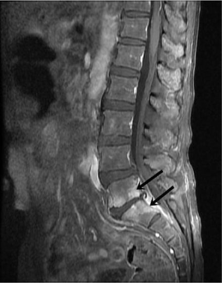
Fig. 1 Spine MRI shows diffuse contrast enhancement of prevertebral soft tissue and endplate at L5-S1 (arrow).
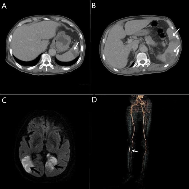
Fig. 2 Septic emboli identified according to imaging modalities (arrow). (A) Contrast-enhanced abdominal CT shows wedge shaped low attenuated lesion at spleen. (B) Aggravated state of spleen after 8 weeks. (C) Brain MRI shows acute ischemic infarct in both occipital lobe. (D) CT angiography of femoral artery shows focal segmental occlusion at distal portion of right popliteal artery.
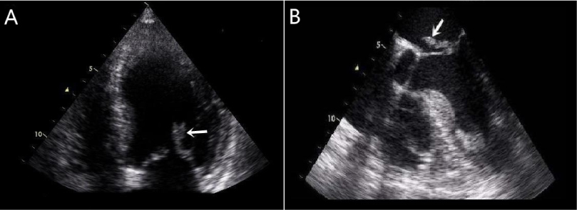
Fig. 3 Vegetation on the posterior leaflet of mitral valve (arrow). (A) Transthoracic echocardiographic finding. (B) Transesophageal echocardiographic finding after 4 weeks.
Reference
-
1. Kim YI, Kim SE, Jang HC, Jung SI, Song SK, Park KH. Analysis of the Clinical Characteristics and Prognostic Factors of Infectious Spondylitis. Infect Chemother. 2011. 43:48–54.
Article2. Koo KH, Lee HJ, Chang BS, Yeom JS, Park KW, Lee CK. Differential Diagnosis between Tuberculous Spondylitis and Pyogenic Spondylitis. J Korean Soc Spine Surg. 2009. 16:112–121.
Article3. Gouliouris T, Aliyu SH, Brown NM. Spondylodiscitis: update on diagnosis and management. J Antimicrob Chemother. 2010. 65:iii11–iii24.
Article4. Murdoch DA. Gram-positive anaerobic cocci. Clin Microbiol Rev. 1998. 11:81–120.
Article5. Zimmerli W. Clinical practice. Vertebral osteomyelitis. N Engl J Med. 2010. 362:1022–1029.6. Cottle L, Riordan T. Infectious spondylodiscitis. J Infect. 2008. 56:401–412.
Article7. Minces LR, Shields RK, Sheridan K, Ho KS, Silveira FP. Peptostreptococcus infective endocarditis and bacteremia. Analysis of cases at tertiary medical center and review of the literature. Anaerobe. 2010. 16:327–330.
Article8. Könönen E, Bryk A, Niemi P, Kanervo-Nordström A. Antimicrobial susceptibilities of peptostreptococcus anaerobius and the newly described peptostreptococcus stomatis isolated from various human sources. Antimicrob Agents Chemother. 2007. 51:2205–2207.
Article9. Lee KY, Chong YS, Jeong SH, Xu XS, Kwon OH. Emerging Resistance of Anaerobic Bacteria to Antimicrobial Agents in South Korea. Clin Infect Dis. 1996. 23:S73–S77.
Article10. The Korean Society of Infectious Diseases. Korean Society for Chemotherapy. The Korean Society of Clinical Microbiology. The Korean Society of Cardiology. The Korean Society for Thoracic and Cardiovascular Surgery. Clinical Guideline for the Diagnosis and Treatment of Cardiovascular Infection. Infect Chemother. 2011. 43:129–177.
- Full Text Links
-
- Actions
-
Cited
- CITED
-
- Close
- Share
-
- Similar articles
-
- Streptococcus Spondylitis Concomitant Infectious Endocarditis: A Case Report
- A Case of Pneumococcal Endocarditis Accompanied by Arthritis and Meningitis
- Clinical Features and Rate of Infective Endocarditis in Non-Faecalis and Non-faecium Enterococcal Bacteremia
- Community-acquired Methicillin-resistant Staphylococcus aureus Bacteremia Complicated by Acute Cholecystitis
- A Case of Infective Endocarditis caused by Abiotrophia defectiva in Korea
