A Butterfly-Shaped Primary Cardiac Lymphoma That Showed Bi-Atrial Involvement
- Affiliations
-
- 1Department of Internal Medicine, The Catholic University of Korea College of Medicine, Seoul, Korea. hhhsungho@hanmail.net
- KMID: 2297907
- DOI: http://doi.org/10.4070/kcj.2012.42.1.46
Abstract
- We described here a patient who presented with symptoms of heart failure who was found to have severe bilateral impairment of atrioventricular inflow. Primary cardiac lymphoma (PCL) with extensive involvement of the two atria, pericardium and myocardium is an extremely rare tumor in immunocompetent patients. We report here a case of PCL in an immunocompetent patient with involvement of both atria and the atrial septum. The tumor had a butterfly shape. We could not do surgical excision because of the massive pericardiac invasion. The diagnosis was B-cell lymphoma and this was confirmed by the pericardiac biopsy.
Keyword
MeSH Terms
Figure
-

Fig. 1 Tranthoracic echocardiography (A) and transesophageal echocardiography (B) showed a 6.5 cm mobile mass in the right atrium and a 3.5 cm mobile mass in the left atrium (arrows). Coronary angiography revealed no stenotic lesions, but we observed feeding vessels (arrows) from the left circumflex artery and the proximal right coronary artery to the primary cardiac lymphoma (C and D).
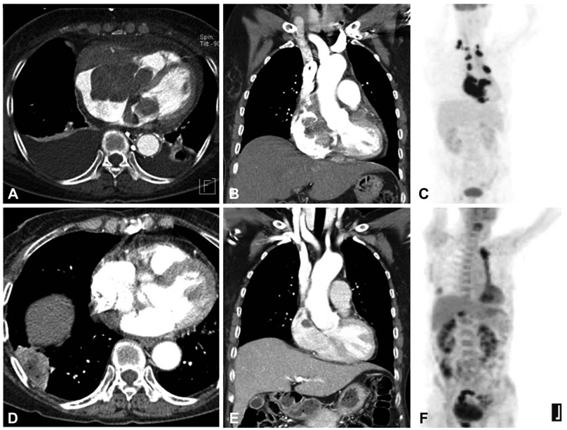
Fig. 2 Cardiac CT (A and B) showed huge bi-atrial masses with invasion into the atrial septum, pericardium and myocardium. Positron emission tomography-CT showed a markedly increased fluorodeoxyglucose uptake in the heart and pericardium and in the mediastinal and right supracalvicular lymph nodes (C). After six rounds of chemotherapy, masses of primary cardiac lymphoma were remarkablely decreased (D, E and F). An Abscess of the right lower pulmonary lobe occurred during chemotherapy (D).
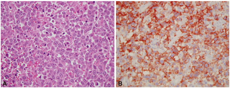
Fig. 3 Hematoxylin-Eosin staining (high power field, ×400) (A) was consistent with a diffuse large B cell lymphoma. This was confirmed by immunohistology. The cells were CD20 B positive (B) and CD3 T negative.
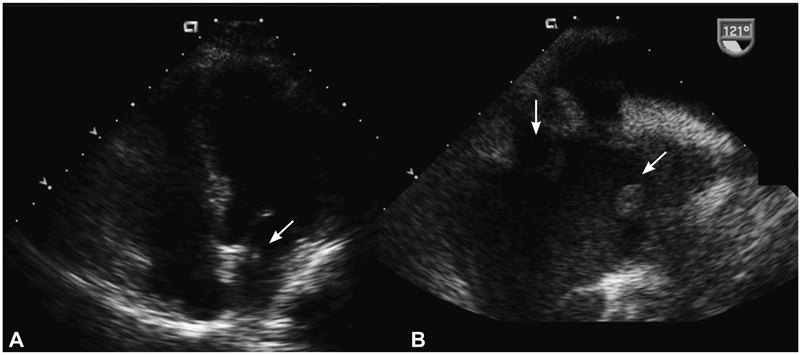
Fig. 4 Follow-up tranthoracic echocardiography (A) and transesophageal echocardiography (B) revealed <1 cm sized remnant masses (arrows) in the both atria.
Reference
-
1. McAllister HA, Fenoglio JJ. Tumors of the cardiovascular system. Atlas of tumor pathology, 2nd series, fascicle 15. 1978. Washington, DC: Armed Forces Institute of Pathology;99–100.2. Lam KY, Dickens P, Chan ACL. Tumors of the heart: a 20-year experience with a review of 12,485 consecutive autopsies. Arch Pathol Lab Med. 1993. 117:1027–1031.3. de Lucas EM, Pagola MA, Fernàndez F, et al. Primary cardiac lymphoma: helical CT findings and radiopathologic correlation. Cardiovasc Intervent Radiol. 2004. 27:190–191.4. Yuh DD, Kubo SH, Francis GS, et al. Primary cardiac lymphoma treated with orthotopic heart transplantation: a case report. J Heart Lung Transplant. 1994. 13:538–542.5. Monsuez JJ, Frija J, Hertz-Pannier L, Miclea JM, Extra JM, Boiron M. Non-Hodgkin's lymphoma with cardiac presentation: evaluation and follow-up with echocardiography and MR imaging. Eur Heart J. 1991. 12:464–467.6. Ikeda H, Nakamura S, Nishimaki H, et al. Primary lymphoma of the heart: case report and literature review. Pathol Int. 2004. 54:187–195.7. Nascimento AF, Winters GL, Pinkus GS. Primary cardiac lymphoma: clinical, histologic, immunophenotypic, and genotypic features of 5 cases of a rare disorder. Am J Surg Pathol. 2007. 31:1344–1350.8. Antoniades L, Eftychiou C, Petrou PM, Bagatzounis A, Minas M. Primary cardiac lymphoma: case report and brief review of the literature. Echocardiography. 2009. 26:214–219.9. Fujita Y, Ikebuchi M, Tarui S, Irie H. Successful combined treatment of primary cardiac malignant lymphoma with urgent cardiac operation and chemotherapy. Circ J. 2009. 73:967–969.10. Chalabreysse L, Berger F, Loire R, Devouassoux G, Cordier JF, Thivolet-Bejui F. Primary cardiac lymphoma in immunocompetent patients: a report of three cases and review of the literature. Virchows Arch. 2002. 441:456–461.11. Somers K, Lothe F. Primary lymphosarcoma of the heart: review of the literature and report of 3 cases. Cancer. 1960. 13:449–457.12. Chao TY, Han SC, Nieh S, Lan GY, Lee SH. Diagnosis of primary cardiac lymphoma. Report of a case with cytologic examination of pericardial fluid and imprints of transvenously biopsied intracardiac tissue. Acta Cytol. 1995. 39:955–959.13. Moore JA, DeRan BP, Minor R, Arthur J, Fraker TD Jr. Transesophageal echocardiographic evaluation of intracardiac lymphoma. Am Heart J. 1992. 124:514–516.14. Jurkovich D, de Marchena E, Bilsker M, Fierro-Renoy C, Temple D, Garcia H. Primary cardiac lymphoma diagnosed by percutaneous intracardiac biopsy with combined fluoroscopic and transesophageal echocardiographic imaging. Catheter Cardiovasc Interv. 2000. 50:226–233.15. Mejhert M, Muller-Suur R. Primary lymphoma of the heart. Scand Cardiovasc J. 2000. 34:606–608.16. Satome M, Yoshitomi Y, Kojima S, Kuramochi M. Primary cardiac lymphoma: a case report. Angiology. 2002. 53:239–242.17. Kim JY, Woo CM, Lee JY, et al. A case of primary cardiac non-Hodgkin's lymphoma. Korean Circ J. 2004. 34:808–812.
- Full Text Links
-
- Actions
-
Cited
- CITED
-
- Close
- Share
-
- Similar articles
-
- Non-Hodgkin’s Lymphoma Presenting as Intracardiac Mass: A Rare Presentation
- Spectral Domain Optical Coherence Tomography Findings of Butterfly Shaped Pigment Dystrophy
- Primary Cardiac Lymphoma: Case Report
- A Case of Cardiac Lymphoma Developed in Right Atrium
- A Case of Primary Nasal CD56+ NK/T cell Lymphoma with Cutaneous Involvement
