Korean J Urol.
2006 Jul;47(7):757-761. 10.4111/kju.2006.47.7.757.
Clinicopathological Significance of the Lymphovascular Invasion Detected in Specimens from Radical Retropubic Prostatectomies
- Affiliations
-
- 1Department of Urology, Seoul National University Bundang Hospital, Seongnam, Korea. selee@snubh.org
- KMID: 2294318
- DOI: http://doi.org/10.4111/kju.2006.47.7.757
Abstract
-
PURPOSE: We tried to determine the clinicopathological significance of lymphovascular invasion (LVI) in patients who were treated for prostate cancer with radical retropubic prostatectomy.
MATERIALS AND METHODS
From November 2003 to June 2005, 165 patients underwent radical retropubic prostatectomy for clinically-localized prostate cancer at our institution. The results of the final pathologic analyses were reviewed.
RESULTS
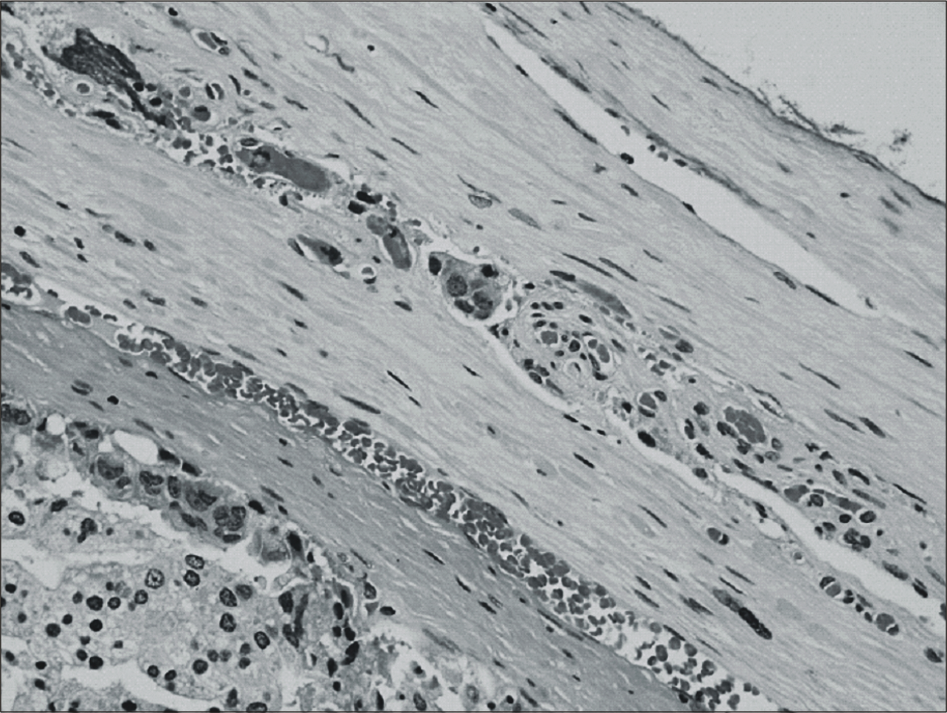
Of the 165 total patients, foci of LVI were identified in 46 patients. LVI was associated with a higher preoperative serum level of prostate-specific antigen (p=0.006), the Gleason score (p<0.0001), a higher weight of tumor volume (p<0.0001), a higher rate of capsular penetration (p<0.0001), a higher rate of seminal vesicle involvement (p<0.0001), and a higher rate of a positive margin (p<0.0001).
CONCLUSIONS
Since the pathological features of LVI appear to be associated with the other established features of more advanced prostate cancers, they may prove to be useful markers for predicting the prognosis of patients who undergo radical prostatectomy. Our findings support performing routine evaluation for LVI in radical prostatectomy specimens and its inclusion in the models for predicting the clinical outcome.
MeSH Terms
Figure
Reference
-
1.Scardino PT. Early detection of prostate cancer. Urol Clin North Am. 1989. 16:635–55.
Article2.Herman CM., Wilcox GE., Kattan MW., Scardino PT., Wheeler TM. Lymphovascular invasion as a predictor of disease progression in prostate cancer. Am J Surg Pathol. 2000. 24:859–63.
Article3.Henson DE., Hutter RV., Farrow G. Practice protocol for the examination of specimens removed from patients with carcinoma of the prostate gland. A publication of the cancer committee, college of American pathologists. Task force on the examination of specimens removed from patients with prostate cancer. Arch Pathol Lab Med. 1994. 118:779–83.4.McNeal JE., Yemoto CE. Significance of demonstrable vascular space invasion for the progression of prostatic adenocarcinoma. Am J Surg Pathol. 1996. 20:1351–60.
Article5.van den Ouden D., Kranse R., Hop WC., van der Kwast TH., Schroder FH. Microvascular invasion in prostate cancer: prognostic significance in patients treated by radical prostatectomy for clinically localized carcinoma. Urol Int. 1998. 60:17–24.
Article6.de la Taille A., Rubin MA., Buttyan R., Olsson CA., Bagiella E., Burchardt M, et al. Is microvascular invasion on radical prostatectomy specimens a useful predictor of PSA recurrence for prostate cancer patients? Eur Urol. 2000. 38:79–84.
Article7.Bahnson RR., Dresner SM., Gooding W., Becich MJ. Incidence and prognostic significance of lymphatic and vascular invasion in radical prostatectomy specimens. Prostate. 1989. 15:149–55.
Article8.Salomao DR., Graham SD., Bostwick DG. Microvascular invasion in prostate cancer correlates with pathologic stage. Arch Pathol Lab Med. 1995. 119:1050–4.9.Bettelheim R., Neville AM. Lymphatic and vascular channel involvement within infiltrative breast carcinoma as a guide to prognosis at the time of primary surgical treatment. Lancet. 1981. 2:631.10.Clemente C., Boracchi P., Andreola S., Del Vecchio M., Veronesi P., Rilke FO. Peritumoral lymphatic invasion in patients with node-negative mammary duct carcinoma. Cancer. 1992. 69:1396–403.
Article11.Gasparini G., Weidner N., Bevilacqua P., Maluta S., Dalla Palma P., Caffo O, et al. Tumor microvessel density, p53 expression, tumor size, and peritumoral lymphatic vessel invasion are relevant prognostic markers in node-negative breast carcinoma. J Clin Oncol. 1994. 12:454–66.
Article12.Lee AK., DeLellis RA., Silverman ML., Wolfe HJ. Lymphatic and blood vessel invasion in breast carcinoma: a useful prognostic indicator? Hum Pathol. 1986. 17:984–7.
Article13.Nime FA., Rosen PP., Thaler HT., Ashikari R., Urban JA. Prognostic significance of tumor emboli in intramammary lymphatics in patients with mammary carcinoma. Am J Surg Pathol. 1977. 1:25–30.
Article14.Edwards JG., Swinson DE., Jones JL., Muller S., Waller DA., O' Byrne KJ. Tumor necrosis correlates with angiogenesis and is a predictor of poor prognosis in malignant mesothelioma. Chest. 2003. 124:1916–23.
- Full Text Links
- Actions
-
Cited
- CITED
-
- Close
- Share
- Similar articles
-
- The Prognostic Significance of Neuroendocrine Differentiation for Treating Prostatic Carcinoma in 699 Cases of Radical Prostatectomy
- Risk Factors for Lymph Node Metastasis and Oncologic Outcomes in Small Rectal Neuroendocrine Tumors with Lymphovascular Invasion
- Inguinal hernia developed after radical retropubic surgery for prostate cancer
- Relationship between CD44v6 Expression and Clinicopathologic Parameters in Endometrial Carcinomas
- Multivariate prognostic analysis of adenocarcinoma of the uterine cervix treated with radical hysterectomy and systematic lymphadenectomy


