Oncoplastic Technique Combining an Adipofascial Flap with an Extended Glandular Flap for the Breast-Conserving Reconstruction of Small Dense Breasts
- Affiliations
-
- 1Department of Breast Surgery, Mie University Graduate School of Medicine, Tsu, Japan. mokomoko@clin.medic.mie-u.ac.jp
- KMID: 2286446
- DOI: http://doi.org/10.4048/jbc.2012.15.4.468
Abstract
- We introduce a method combining two oncoplastic techniques for breast-conserving reconstruction. The procedure is as follows: first, an extended glandular flap is made by undermining the breast from both the skin and the pectoralis fascia to the upper edge of the breast at the subclavicular area. After modeling the breast mound with the extended glandular flap, an inframammary adipofascial flap is made. The flap is reflected back to the breast area remodeled using the extended glandular flap. After reshaping the breast, the inframammary line is then re-shaped. This method is indicated for patients with breast cancer in the outer portion of the breast, who have small dense breasts, and have undergone a large excision of about 40% of their breast volume. We treated four patients, all of whom had either excellent or good cosmetic results with no fat necrosis.
MeSH Terms
Figure
-
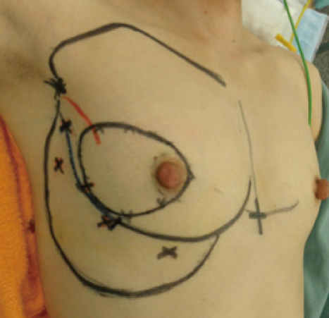
Figure 1 The design before the operation. Marking the partial resection area, the upper edge of the breast at the subclavicular area, and the position of the nipple in the standing position, as well as the inframammary groove, the area of the adipofascial flap, and the location of perforators (x mark).
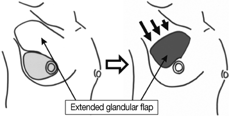
Figure 2 The extended glandular flap. The extended glandular flap is made by freeing the breast from both the skin and the pectoralis fascia. The flap is moved into the defect in the direction of arrows, and the breast mound is remodeled.
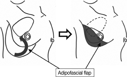
Figure 3 The inframammary adipofascial flap. The inframammary adipofascial flap is reflected back to the breast area (arrow) that was remodeled by using the extended glandular flap.
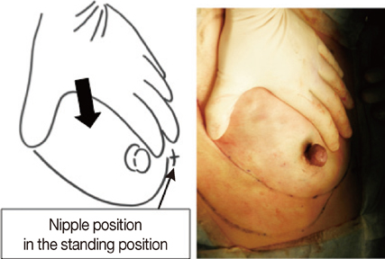
Figure 4 Checking the shape of the breast. The nipple position is set at the position that had been marked before surgery with the patient in the standing position while applying pressure from the upper side (arrow).

Figure 5 Photographs of case 1, showing the good cosmetic results. (A, B) Preoperative photographs: (A) frontal view, (B) oblique view. (C, D) Postoperative photographs taken 6.6 years after the operation: (C) frontal view, (D) oblique view.
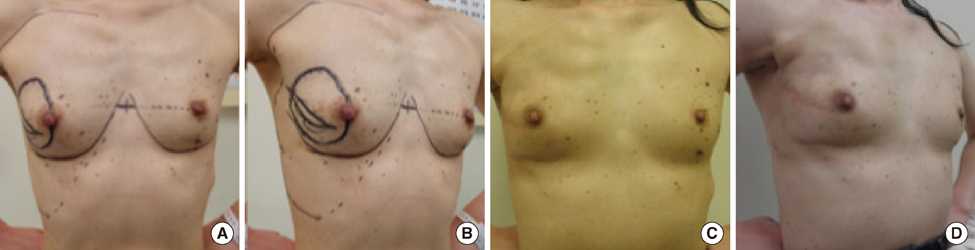
Figure 6 Photographs of case 2, showing the excellent cosmetic results. (A, B) Preoperative photographs: (A) frontal view, (B) oblique view. (C, D) Postoperative photographs taken 3.1 years after the operation: (C) frontal view, (D) oblique view.
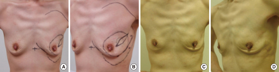
Figure 7 Photographs of case 3, showing the excellent cosmetic results. (A, B) Preoperative photographs: (A) frontal view, (B) oblique view. (C, D) Postoperative photographs taken 2.4 years after the operation: (C) frontal view, (D) oblique view.

Figure 8 Photographs of case 4, showing the good cosmetic results. (A, B) Preoperative photographs: (A) frontal view, (B) oblique view. (C, D) Postoperative photographs taken 1.3 years after the operation: (C) frontal view, (D) oblique view.
Reference
-
1. Bulstrode NW, Shrotria S. Prediction of cosmetic outcome following conservative breast surgery using breast volume measurements. Breast. 2001. 10:124–126.
Article2. Clough KB, Kaufman GJ, Nos C, Buccimazza I, Sarfati IM. Improving breast cancer surgery: a classification and quadrant per quadrant atlas for oncoplastic surgery. Ann Surg Oncol. 2010. 17:1375–1391.
Article3. Ogawa T, Hanamura N, Yamashita M, Ri Y, Kuriyama N, Isaji S. Usefulness of breast-volume replacement using an inframammary adipofascial flap after breast-conservation therapy. Am J Surg. 2007. 193:514–518.
Article4. Ogawa T, Hanamura N, Yamashita M, Kimura H, Kashikura Y. Long-term results of breast volume replacement using an inframammary adipofascial flap after breast-conserving surgery. Breast Cancer. In press.
Article5. Ogawa T, Hanamura N, Yamashita M, Kimura H, Kashikura Y. Breast-volume displacement using an extended glandular flap for small dense breasts. Plast Surg Int. 2011. 2011:359842.
Article6. Noguchi M, Taniya T, Miyazaki I, Saito Y. Immediate transposition of a latissimus dorsi muscle for correcting a postquadrantectomy breast deformity in Japanese patients. Int Surg. 1990. 75:166–170.7. Rainsbury RM, Paramanathan N. Recent progress with breast-conserving volume replacement using latissimus dorsi miniflaps in UK patients. Breast Cancer. 1998. 5:139–147.
Article8. Hamdi M, Spano A, Van Landuyt K, D'Herde K, Blondeel P, Monstrey S. The lateral intercostal artery perforators: anatomical study and clinical application in breast surgery. Plast Reconstr Surg. 2008. 121:389–396.
Article9. Hamdi M, Van Landuyt K, Hijjawi JB, Roche N, Blondeel P, Monstrey S. Surgical technique in pedicled thoracodorsal artery perforator flaps: a clinical experience with 99 patients. Plast Reconstr Surg. 2008. 121:1632–1641.
Article10. Harris JR, Levene MB, Svensson G, Hellman S. Analysis of cosmetic results following primary radiation therapy for stages I and II carcinoma of the breast. Int J Radiat Oncol Biol Phys. 1979. 5:257–261.
Article11. Kronowitz SJ, Hunt KK, Kuerer HM, Strom EA, Buchholz TA, Ensor JE, et al. Practical guidelines for repair of partial mastectomy defects using the breast reduction technique in patients undergoing breast conservation therapy. Plast Reconstr Surg. 2007. 120:1755–1768.
Article12. McCulley SJ, Macmillan RD. Planning and use of therapeutic mammoplasty: Nottingham approach. Br J Plast Surg. 2005. 58:889–901.
- Full Text Links
-
- Actions
-
Cited
- CITED
-
- Close
- Share
-
- Similar articles
-
- Surgical Techniques for Personalized Oncoplastic Surgery in Breast Cancer Patients with Small- to Moderate-Sized Breasts (Part 2): Volume Replacement
- Usefulness of Oncoplastic Volume Replacement Techniques after Breast Conserving Surgery in Small to Moderate-sized Breasts
- Oncoplastic Breast Surgery
- The usefulness of pedicled perforator flap in partial breast reconstruction after breast conserving surgery in Korean women
- Oncoplastic Surgical Techniques for Personalized Breast Conserving Surgery in Breast Cancer Patient with Small to Moderate Sized Breast
