J Breast Cancer.
2015 Mar;18(1):44-49. 10.4048/jbc.2015.18.1.44.
Ultrasonography-Guided Surgical Clip Placement for Tumor Localization in Patients Undergoing Neoadjuvant Chemotherapy for Breast Cancer
- Affiliations
-
- 1Department of Radiology, Kangbuk Samsung Hospital, Sungkyunkwan University School of Medicine, Seoul, Korea. seonhyeong.choi@samsung.com
- 2Department of Surgery, Kangbuk Samsung Hospital, Sungkyunkwan University School of Medicine, Seoul, Korea.
- 3Department of Pathology, Kangbuk Samsung Hospital, Sungkyunkwan University School of Medicine, Seoul, Korea.
- KMID: 2286336
- DOI: http://doi.org/10.4048/jbc.2015.18.1.44
Abstract
- PURPOSE
We investigated the feasibility of using surgical clips as markers for tumor localization and their effect on the imaging evaluation of treatment responses after neoadjuvant chemotherapy (NAC).
METHODS
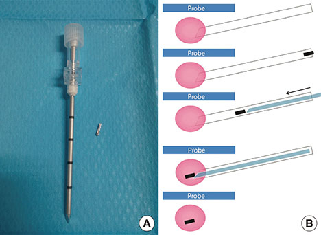
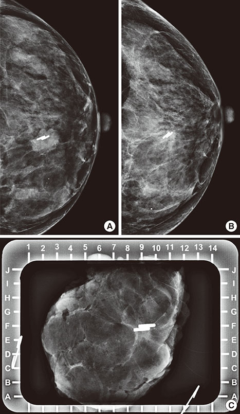
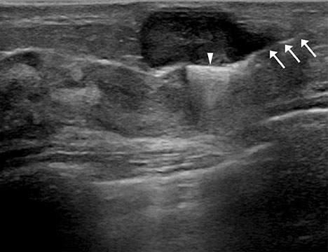
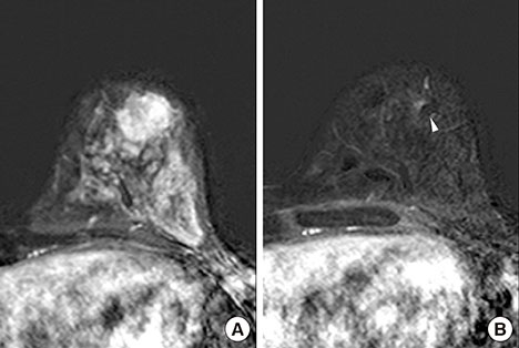
A total of 16 breast cancers confirmed by needle biopsy in 15 patients were included in this study from October 2012 to June 2014. Under ultrasonography (US)-guidance, the surgical clips were placed prior to NAC. Additional mammography, breast US, and breast magnetic resonance examinations were performed within 10 days before surgery. The time period from marker insertion to operation date was documented. Images acquired via the three modalities were evalu-ated for the following parameters: location of clip, clip migration (>1 cm), the presence of complications from clip placement, and the effect of clips on the assessment of treatment.
RESULTS
The mean time period was 128.6+/-34.4 days (median, 132.0 days) from the date of clip insertion to the date of surgery. The mean number of inserted clips was 2.3+/-0.7 (median, 2.0). Clip migration was not visualized by imaging in any patient, and there were no complications reported. Surgical clips did not negatively affect the assessment of treatment responses to NAC.
CONCLUSION
Surgical clips may replace commercial tissue markers for tumor localization in breast cancer patients undergoing NAC without migration. Surgical clips are well tolerated and safe for the patient, easily visualized on imaging, do not interfere with treatment response, and are cost-effective.
Keyword
MeSH Terms
Figure
Cited by 1 articles
-
Pathologic Evaluation of Breast Cancer after Neoadjuvant Therapy
Cheol Keun Park, Woo-Hee Jung, Ja Seung Koo
J Pathol Transl Med. 2016;50(3):173-180. doi: 10.4132/jptm.2016.02.02.
Reference
-
1. Kaufmann M, von Minckwitz G, Bear HD, Buzdar A, McGale P, Bonnefoi H, et al. Recommendations from an international expert panel on the use of neoadjuvant (primary) systemic treatment of operable breast cancer: new perspectives 2006. Ann Oncol. 2007; 18:1927–1934.
Article2. Oh JL, Nguyen G, Whitman GJ, Hunt KK, Yu TK, Woodward WA, et al. Placement of radiopaque clips for tumor localization in patients undergoing neoadjuvant chemotherapy and breast conservation therapy. Cancer. 2007; 110:2420–2427.
Article3. Abdel-Razeq H, Marei L. Current neoadjuvant treatment options for HER2-positive breast cancer. Biologics. 2011; 5:87–94.4. Kim Z, Min SY, Yoon CS, Lee HJ, Lee JS, Youn HJ, et al. The basic facts of Korean breast cancer in 2011: results of a nationwide survey and breast cancer registry database. J Breast Cancer. 2014; 17:99–106.
Article5. Lobbes MB, Prevos R, Smidt M, Tjan-Heijnen VC, van Goethem M, Schipper R, et al. The role of magnetic resonance imaging in assessing residual disease and pathologic complete response in breast cancer patients receiving neoadjuvant chemotherapy: a systematic review. Insights Imaging. 2013; 4:163–175.
Article6. Dash N, Chafin SH, Johnson RR, Contractor FM. Usefulness of tissue marker clips in patients undergoing neoadjuvant chemotherapy for breast cancer. AJR Am J Roentgenol. 1999; 173:911–917.
Article7. Kaufmann M, von Minckwitz G, Mamounas EP, Cameron D, Carey LA, Cristofanilli M, et al. Recommendations from an international consensus conference on the current status and future of neoadjuvant systemic therapy in primary breast cancer. Ann Surg Oncol. 2012; 19:1508–1516.
Article8. Margolin FR, Jacobs RP, Denny SR, Schrumpf JD. Clip placement after sonographically guided percutaneous breast biopsy. Breast J. 2003; 9:226–230.
Article9. Kopans DB. Clip placement during sonographically guided breast biopsy. AJR Am J Roentgenol. 2001; 176:1076–1077.
Article10. Guenin MA. Clip placement during sonographically guided large-core breast biopsy for mammographic-sonographic correlation. AJR Am J Roentgenol. 2000; 175:1053–1055.
Article11. Phillips SW, Gabriel H, Comstock CE, Venta LA. Sonographically guided metallic clip placement after core needle biopsy of the breast. AJR Am J Roentgenol. 2000; 175:1353–1355.
Article12. Genson CC, Blane CE, Helvie MA, Waits SA, Chenevert TL. Effects on breast MRI of artifacts caused by metallic tissue marker clips. AJR Am J Roentgenol. 2007; 188:372–376.
Article13. Tamai K, Mitsumori M, Fujishiro S, Kokubo M, Ooya N, Nagata Y, et al. A case of allergic reaction to surgical metal clips inserted for postoperative boost irradiation in a patient undergoing breast-conserving therapy. Breast Cancer. 2001; 8:90–92.
Article14. Coles CE, Wilson CB, Cumming J, Benson JR, Forouhi P, Wilkinson JS, et al. Titanium clip placement to allow accurate tumour bed localization following breast conserving surgery: audit on behalf of the IMPORT Trial Management Group. Eur J Surg Oncol. 2009; 35:578–582.
Article15. Uematsu T. Commercially available titanium clip placement following a sonographically guided core needle biopsy of the breast. Breast J. 2007; 13:624–626.
Article16. Lee SY, Kook SH, Kwag HJ. The results and usefulness of marker clip placement after ultrasound-guided mammotome excision of breast lesion. J Korean Radiol Soc. 2005; 52:207–213.
Article17. Margolin FR, Kaufman L, Denny SR, Jacobs RP, Schrumpf JD. Metallic marker placement after stereotactic core biopsy of breast calcifications: comparison of two clips and deployment techniques. AJR Am J Roentgenol. 2003; 181:1685–1690.
Article18. Baron LF, Baron PL, Ackerman SJ, Durden DD, Pope TL Jr. Sonographically guided clip placement facilitates localization of breast cancer after neoadjuvant chemotherapy. AJR Am J Roentgenol. 2000; 174:539–540.
Article
- Full Text Links
- Actions
-
Cited
- CITED
-
- Close
- Share
- Similar articles
-
- US-guided Clip Implantation for Tumor Localization in Breast Cancer Patients Who Undergo Neoadjuvant Chemotherapy: Feasibility Study
- Comparison of breast tissue markers for tumor localization in breast cancer patients undergoing neoadjuvant chemotherapy
- Feasibility of Ultrasound-Guided Localization for Clipped Metastatic Axillary Lymph Nodes After Neoadjuvant Chemotherapy in Breast Cancer Patients: A Pilot Study
- Ultrasonography-Guided Breast Tissue Marker Insertion
- The Results and Usefulness of Marker Clip Placement after Ultrasound-guided Mammotome Excision of Breast Lesion


