Korean J Pediatr Gastroenterol Nutr.  2011 Jun;14(2):130-136. 10.5223/kjpgn.2011.14.2.130.

Plain Abdominal Radiography in Infants and Children

Affiliations
  • 1Department of Radiology, Keimyung University School of Medicine, Daegu, Korea. hjlee@dsmc.or.kr

Abstract

Plain X-ray radiographs are the first line of investigation taken in the diagnosis of abdominal pathology and are considered an important diagnostic tool to provide guidelines for further imaging studies and comprehensive therapeutic management. Although most abdominal pathology demonstrates non-specific radiologic findings, the plain abdominal radiography is very useful in specific diseases, including certain gastrointestinal anomalies. This review provides image findings of normal plain abdominal radiography and some common abdominal pathology in infants and children.

Keyword

Radiography; Abdominal; X-rays; Infant; Newborn; Child

MeSH Terms

Child
Humans
Infant
Infant, Newborn
Radiography, Abdominal

Figure

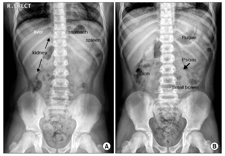
  • Fig. 1 Erect (A) and supine (B) views of the abdominal radiography in a normal child (thin arrows: kidney, thick arrow: psoas shadow).

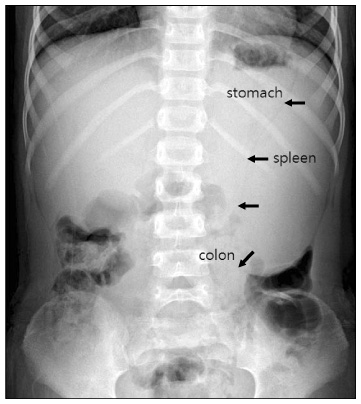
  • Fig. 2 Splenomegaly. The enlarging spleen (arrows) in the left upper quadrant displaces the splenic flexure of the colon caudally and medially, and the stomach medially.

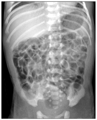
  • Fig. 3 Supine view of a normal neonate. The gas distribution is present throughout the small and large bowel and is one of multiple, closely apposed, rounded or polyhedral structures. The small and large bowel cannot be distinguished.

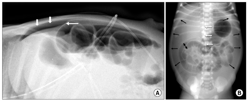
  • Fig. 4 Pneumoperitoneum. The gas is under the anterior abdominal wall (short arrows) and the ligamentum falcifarum is demonstrated (long white arrow) on a cross table lateral view (A). Supine view (B) shows a large oval air collection (long black arrows) in the central abdomen with the shape of a 'football'. It may be identified by the presence of air on both sides of the bowel wall (short black arrows) or air outlining the ligamentum falcifarum (long white arrows).

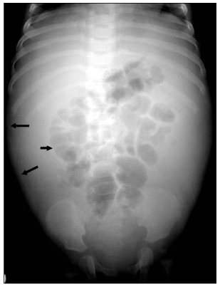
  • Fig. 5 Ascites in a neonate with meconium peritonitis. Supine view demonstrates separation of the flank fat stripe (long arrows) from gas in the descending colon (short arrow) and central displacement of bowel loops.

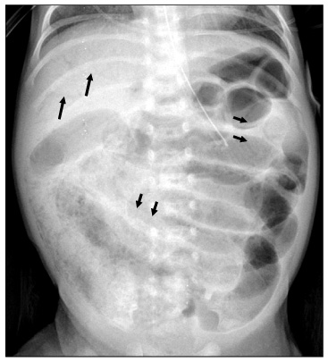
  • Fig. 6 Necrotizing enterocolitis. Supine view shows diffusely dilated bowels with linear intramural gases (short arrows) (pneumatosis cystoides intestinalis) and also multiple linear and branching airs (long arrows) in the liver (air-portogram).

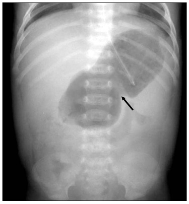
  • Fig. 7 Hypertrophic pyloric stenosis. Supine view demonstrates over distension of the stomach and evidence of a peristaltic wave (arrow) along the gastric body. Note the relatively sparse bowel gas.

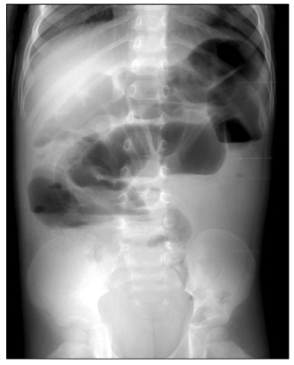
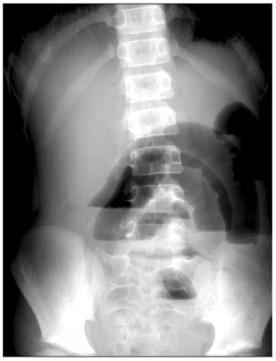
  • Fig. 8 Adynamic ileus with gastroenteritis. The fluid levels tend to be long and are the same level on an erect view. Note gases in both colon and small bowel loops.

  • Fig. 9 Mechanical small bowel obstruction. Erect view shows absent colon gas and multiple different air-fluid levels in the dilated small bowel loop with a 'ladder' appearance.

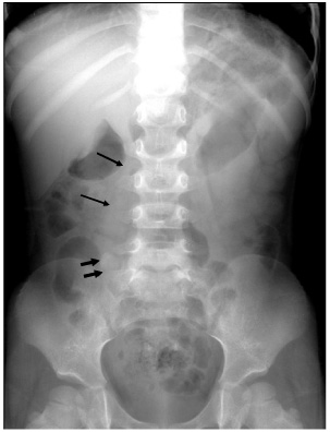
  • Fig. 10 Appendicitis with an appendicolith. Supine view illustrates the presence of a faecolith in the right lower quadrant (short arrows) and loss of the right psoas outline (long arrows). Note normal well-defined left psoas shadow.

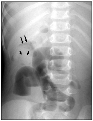
  • Fig. 11 Intussusception with the 'target' and 'meniscus' signs. Supine view shows a round soft-tissue mass in the right upper quadrant. The mass contains a ring-like area of lucency representing the 'target' sign (small arrows). The mass protrudes into the gas-filled transverse colon representing the 'meniscus' sign (long arrows).


Reference

1. Coley BD, Hernans-Schulman M. Slovis TL, editor. The abdomen, pelvis, retroperitoneum. Caffey's pediatric diagnostic imaging. 2008. 11th ed. Philladelphia: Mosby;1759–2464.
2. String DA. Carty H, Brunelle F, Stringer DA, Kao SCS, editors. The gastrointestinal tract. Imaging children. 2005. 2nd ed. Philadelphia: Elsevier Ltd;1289–1529.
3. Swichuk LE. Imaging of the newborn, infant, and young child. 2004. 5th ed. Philadelphia: Lippncott Williams and Wilkins;539–589.
4. McAlister WH, Kronemer KA. Emergency gastrointestinal radiology of the newborn. Radiol Clin North Am. 1996. 34:819–844.
5. Silva AC, Pimenta M, Guimaraes LS. Small bowel obstruction: what to look for. Radiographics. 2009. 29:423–439.
Article
6. Del-Pozo G, Albillos JC, Tejedor D, Calero R, Miguel Rasero M, de-la-Calle U, et al. Intussusception in children: current concepts in diagnosis and enema reduction. Radiographics. 1999. 19:299–319.
Article
Full Text Links
  • KJPGN
Actions
Cited
CITED
export Copy
Close
Share
  • Twitter
  • Facebook
Similar articles
Copyright © 2024 by Korean Association of Medical Journal Editors. All rights reserved.     E-mail: koreamed@kamje.or.kr