Three dimensional analysis of tooth movement using different types of maxillary molar distalization appliances
- Affiliations
-
- 1Division of Orthodontics, Department of Dentistry, College of Medicine, Ewha Womans University, Korea. yschun@ewha.ac.kr
- 2Department of Preventive Medicine, College of Medicine, Ewha Womans University, Korea.
- KMID: 2273969
- DOI: http://doi.org/10.4041/kjod.2008.38.6.376
Abstract
OBJECTIVE
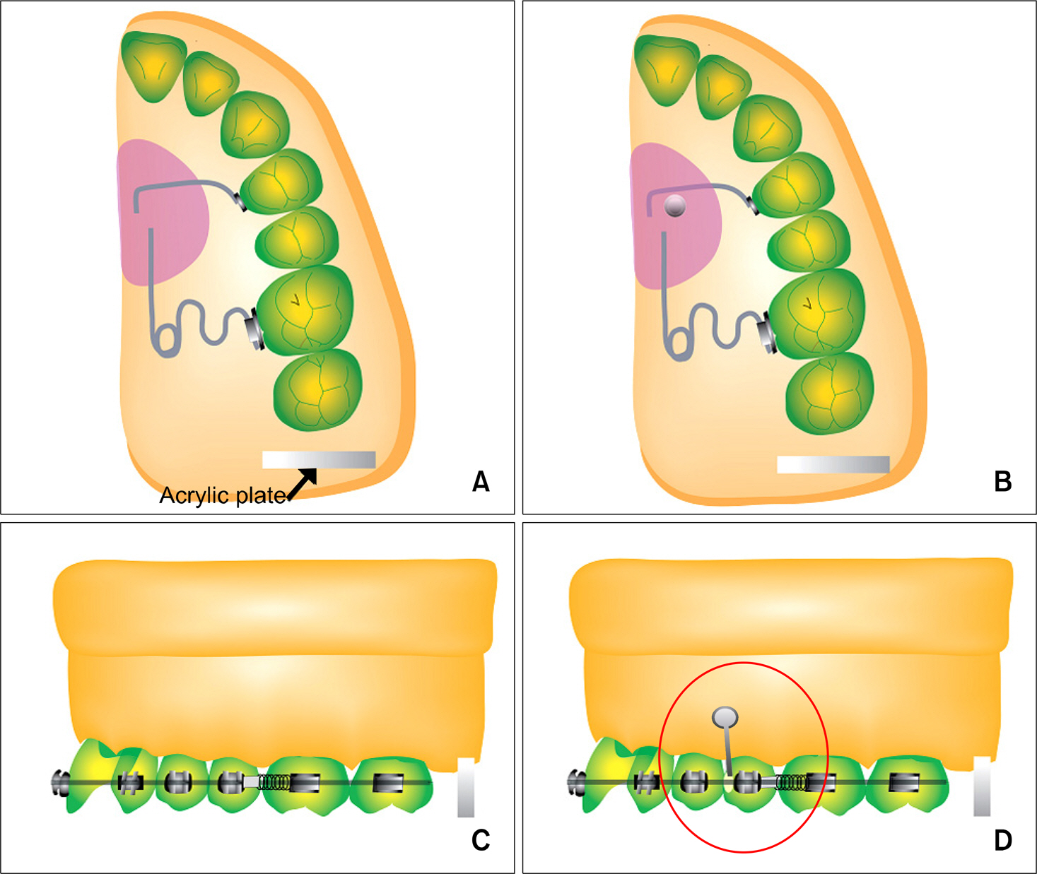
The purpose of this study was to compare the three dimensional changes of tooth movement using four different types of maxillary molar distalization appliances; pendulum appliance (PD), mini-implant supported pendulum appliance (MPD), stainless steel open coil spring (SP) and mini-implant supported stainless steel open coil spring (MSP).
METHODS
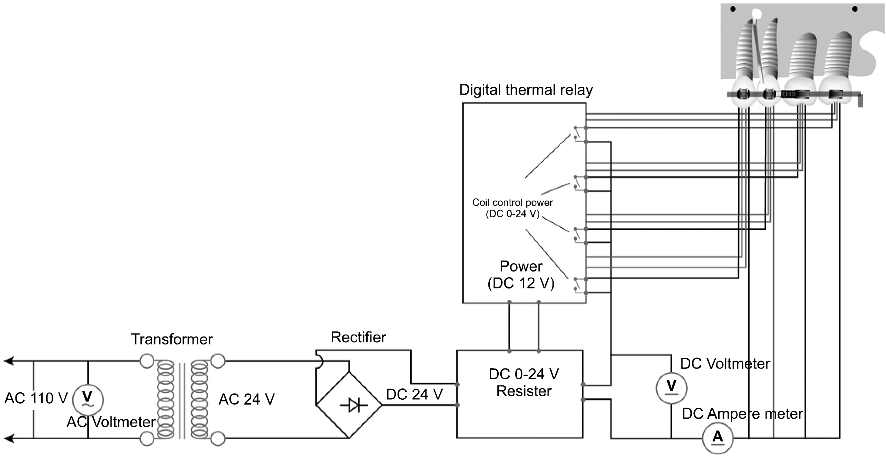
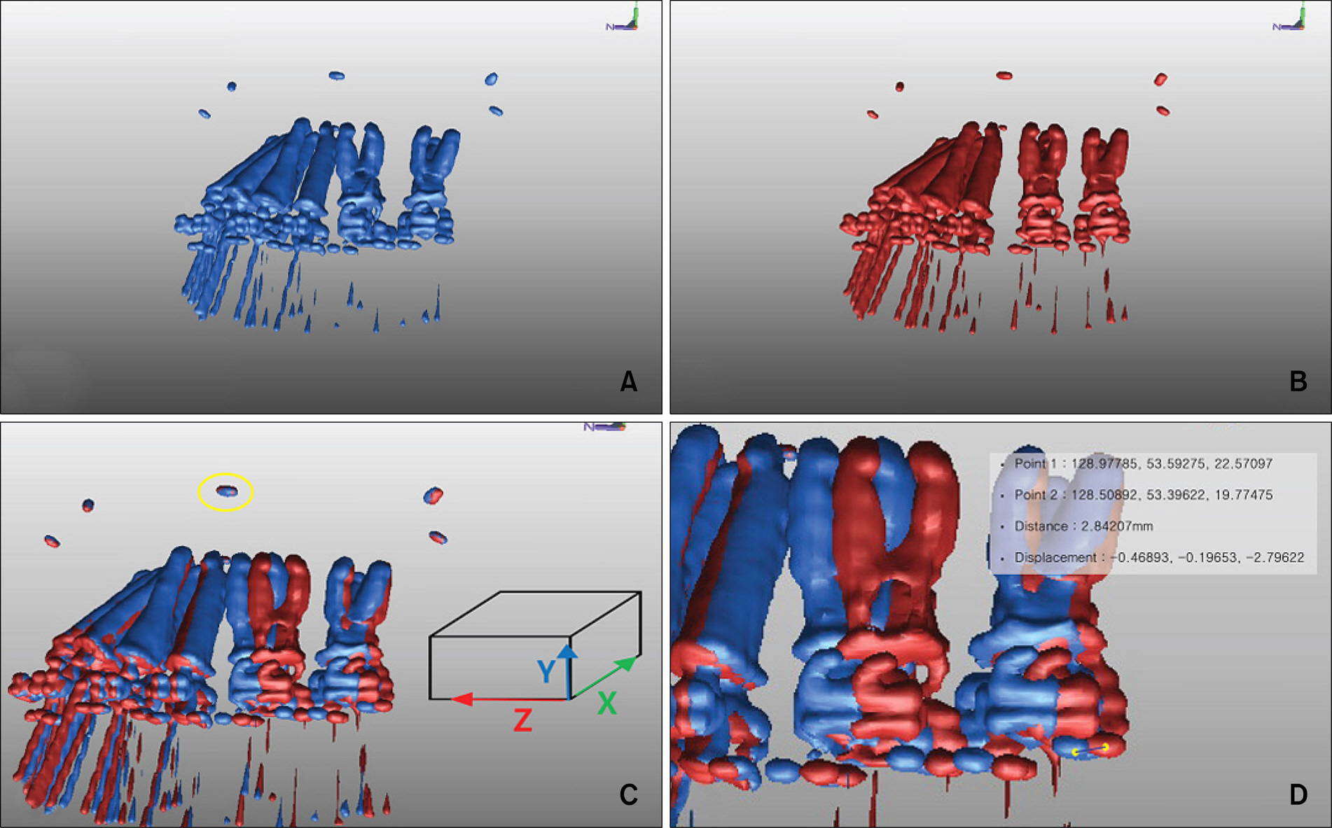
These experiments were performed using the Calorific machine? which can simulate dynamic tooth movement. Computed tomography (CT) images of the experimental model were taken before and after tooth movement in 1 mm thicknesses and reconstructed into a three dimensional model using V-works 4.0TM. These reconstructed images were superimposed using Rapidform 2004TM and the direction and amount of tooth movement were measured.
RESULTS
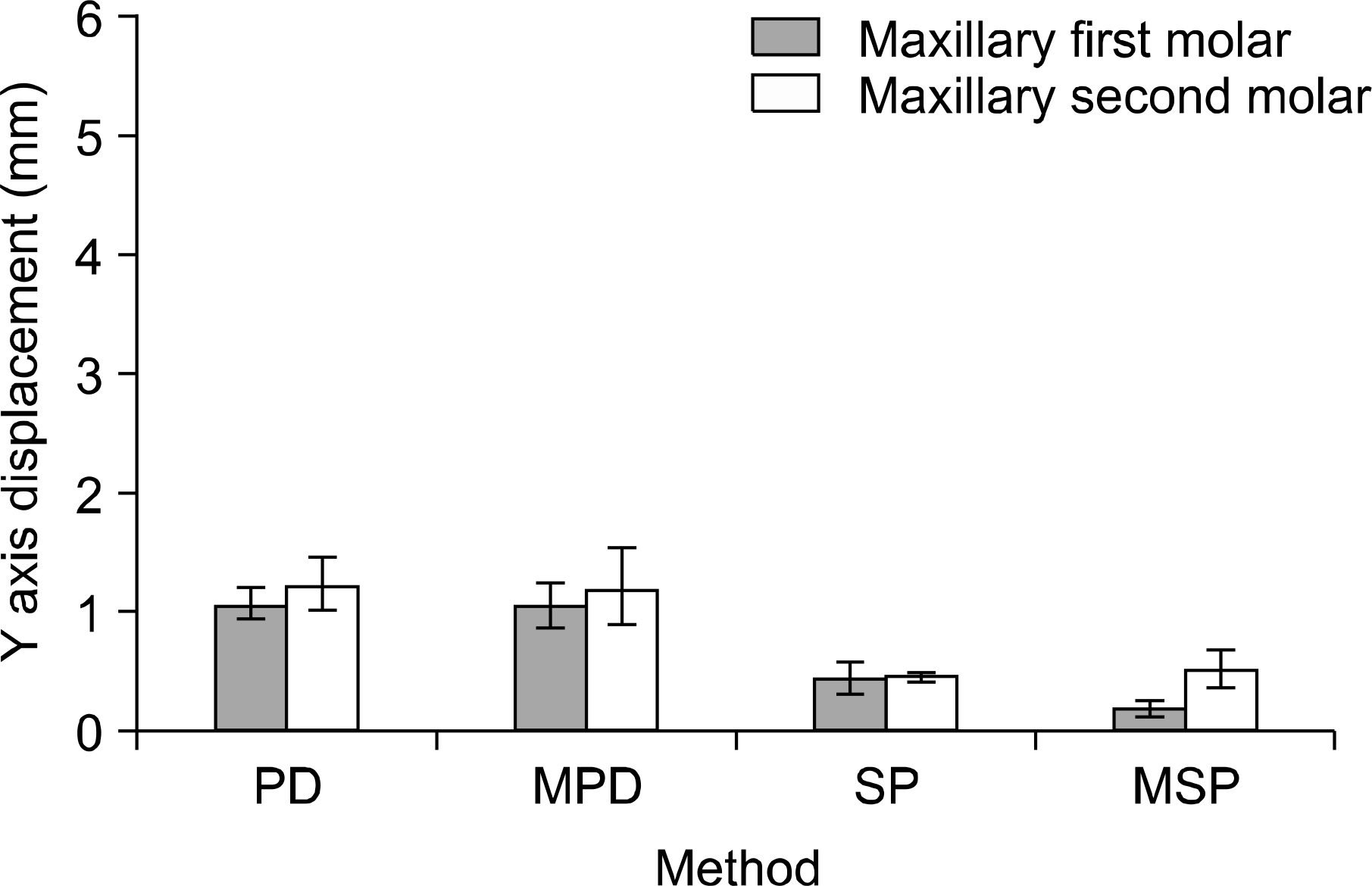
The mean reciprocal anchor loss ratio at the first premolar was 17 - 19% for the PD and SP groups. The appliances using mini-implants (MPD or MSP) resulted in less anchorage loss (7 - 8%). On application of a pendulum appliance or MPD, distalization was obtained by tipping rather than by bodily movement. Furthermore, the maxillary second molar tipped distally and bucally. But on application of MSP, distalization was achieved almost by bodily movement.
CONCLUSIONS
Regarding tooth movement patterns during molar distalization, stainless steel open coil spring with indirect skeletal anchorage was relatively superior to other methods.
Figure
Cited by 6 articles
-
Finite-element investigation of the center of resistance of the maxillary dentition
Gwang-Mo Jeong, Sang-Jin Sung, Kee-Joon Lee, Youn-Sic Chun, Sung-Seo Mo
Korean J Orthod. 2009;39(2):83-94. doi: 10.4041/kjod.2009.39.2.83.The frog appliance for upper molar distalization: a case report
Mehmet Bayram, Metin Nur, Dogan Kilkis
Korean J Orthod. 2010;40(1):50-60. doi: 10.4041/kjod.2010.40.1.50.A reliable method for evaluating upper molar distalization: Superimposition of three-dimensional digital models
Ruhi Nalcaci, Ayse Burcu Kocoglu-Altan, Ali Altug Bicakci, Firat Ozturk, Hasan Babacan
Korean J Orthod. 2015;45(2):82-88. doi: 10.4041/kjod.2015.45.2.82.Finite element analysis of maxillary incisor displacement during en-masse retraction according to orthodontic mini-implant position
Jae-Won Song, Joong-Ki Lim, Kee-Joon Lee, Sang-Jin Sung, Youn-Sic Chun, Sung-Seo Mo
Korean J Orthod. 2016;46(4):242-252. doi: 10.4041/kjod.2016.46.4.242.Effects of bracket slot size during
en-masse retraction of the six maxillary anterior teeth using an induction-heating typodont simulation system
Ji-Yong Kim, Won-Jae Yu, Prasad N. K. Koteswaracc, Hee-Moon Kyung
Korean J Orthod. 2017;47(3):158-166. doi: 10.4041/kjod.2017.47.3.158.Zygoma-gear appliance for intraoral upper molar distalization
Metin Nur, Mehmet Bayram, Alper Pampu
Korean J Orthod. 2010;40(3):195-206. doi: 10.4041/kjod.2010.40.3.195.
Reference
-
1.Gianelly AA., Vaitas AS., Thomas WM., Berger DG. Distalization of molars with repelling magnets. J Clin Orthod. 1988. 22:40–4.2.Gianelly AA., Vaitas AS., Thomas WM. The use of magnets to move molars distally. Am J Orthod Dentofacial Orthop. 1989. 96:161–7.
Article3.Hilgers JJ. The pendulum appliance for Class II non-compliance therapy. J Clin Orthod. 1992. 26:706–14.4.Gianelly AA., Bednar J., Dietz VS. Japanese NiTi coils used to move molars distally. Am J Orthod Dentofacial Orthop. 1991. 99:564–6.
Article5.Jones RD., White JM. Rapid Class II molar correction with an open-coil jig. J Clin Orthod. 1992. 26:661–4.6.Carano A., Testa M. The distal jet for upper molar distalization. J Clin Orthod. 1996. 30:374–80.7.Wilson WL. Variations of labiolingual therapy in Class II cases. Am J Orthod. 1955. 41:852–71.
Article8.Karlsson I., Bondemark L. Intraoral maxillary molar distalization. Angle Orthod. 2006. 76:923–9.
Article9.Pieringer M., Droschl H., Permann R. Distalization with a Nance appliance and coil springs. J Clin Orthod. 1997. 31:321–6.10.Kircelli BH., Pektas ZO., Kircelli C. Maxillary molar distalization with a bone-anchored pendulum appliance. Angle Orthod. 2006. 76:650–9.11.Chang YJ., Lee HS., Chun YS. Microscrew anchorage for molar intrusion. J Clin Orthod. 2004. 38:325–30.12.Yun SW., Lim WH., Chun YS. Molar control using indirect miniscrew anchorage. J Clin Orthod. 2005. 39:661–4.13.Weijs WA., de Jongh HJ. Strain in mandibular alveolar bone during mastication in the rabbit. Arch Oral Biol. 1977. 22:667–75.
Article14.Burstone CJ., Pryputniewicz RJ. Holographic determination of centers of rotation produced by orthodontic forces. Am J Orthod. 1980. 77:396–409.
Article15.Caputo AA., Chaconas SJ., Hayashi RK. Photoelastic visualization of orthodontic forces during canine retraction. Am J Orthod. 1974. 65:250–9.
Article16.Baeten LR. Canine retraction: a photoelastic study. Am J Orthod. 1975. 67:11–23.
Article17.Moss ML., Skalak R., Patel H., Sen K., Moss-Salentijn L., Shinozuka M, et al. Finite element method modeling of craniofacial growth. Am J Orthod. 1985. 87:453–72.
Article18.Ogura M., Yamagata K., Kubota S., Kim JH., Kuroe K., Ito G. Comparison of tooth movements using Friction-Free and preadjusted edgewise bracket systems. J Clin Orthod. 1996. 30:325–30.19.Drescher D., Bourauel C., Thier M. Application of the orthodontic measurement and simulation system (OMSS) in orthodontics. Eur J Orthod. 1991. 13:169–78.
Article20.Rhee JN., Chun YS., Row J. A comparison between friction and frictionless mechanics with a new typodont simulation system. Am J Orthod Dentofacial Orthop. 2001. 119:292–9.
Article21.Chun YS., Row J., Suh MS., Park IK. An experimental study on the dynamic tooth moving effects of two precision lingual archs (Pla) for correction posterior scissor bite by the calorific machine. Korean J Orthod. 1998. 28:29–41.22.Ghosh J., Nanda RS. Evaluation of an intraoral maxillary molar distalization technique. Am J Orthod Dentofacial Orthop. 1996. 110:639–46.
Article23.KeleşA. Erverdi N., Sezen S. Bodily distalization of molars with absolute anchorage. Angle Orthod. 2003. 73:471–82.24.Wehrbein H., Feifel H., Diedrich P. Palatal implant anchorage reinforcement of posterior teeth: a prospective study. Am J Orthod Dentofacial Orthop. 1999. 116:678–86.
Article25.Carano A., Velo S., Leone P., Siciliani G. Clinical applications of the Miniscrew Anchorage System. J Clin Orthod. 2005. 39:9–24.26.Deguchi T., Takano-Yamamoto T., Kanomi R., Hartsfield JK Jr., Roberts WE., Garetto LP. The use of small titanium screws for orthodontic anchorage. J Dent Res. 2003. 82:377–81.27.Sugawara J., Kanzaki R., Takahashi I., Nagasaka H., Nanda R. Distal movement of maxillary molars in nongrowing patients with the skeletal anchorage system. Am J Orthod Dentofacial Orthop. 2006. 129:723–33.
Article28.Chang HN., Hsiao HY., Tsai CM., Roberts WE. Bone-screw anchorage for pendulum appliances and other fixed mechanics applications. Semin Orthod. 2006. 12:284–9.
Article29.Byloff FK., Darendeliler MA. Distal molar movement using the pendulum appliance. Part 1: clinical and radiological evaluation. Angle Orthod. 1997. 67:249–60.30.Bondemark L., Kurol J. Class II correction with magnets and superelastic coils followed by straight-wire mechanotherapy. Occlusal changes during and after dental therapy. J Orofac Orthop. 1998. 59:127–38.31.Bondemark L., Kurol J., Bernhold M. Repelling magnets versus superelastic nickel-titanium coils in simultaneous distal movement of maxillary first and second molars. Angle Orthod. 1994. 64:189–98.32.Chaconas SJ., Caputo AA., Harvey K. Orthodontic force characteristics of open coil springs. Am J Orthod. 1984. 85:494–7.
Article
- Full Text Links
- Actions
-
Cited
- CITED
-
- Close
- Share
- Similar articles
-
- Analysis of midpalatal miniscrew-assisted maxillary molar distalization patterns with simultaneous use of fixed appliances: A preliminary study
- Combined treatment with headgear and the Frog appliance for maxillary molar distalization: a randomized controlled trial
- Maxillary molar derotation and distalization by using a nickel-titanium wire fabricated on a setup model
- Evaluation of the effects of the third molar on distalization and the effects of attachments on distalization and expansion with clear aligners: Three-dimensional finite element study
- Zygoma-gear appliance for intraoral upper molar distalization


