Case series of precursor B-cell lymphoblastic lymphoma
- Affiliations
-
- 1Department of Internal Medicine, Asan Medical Center, University of Ulsan College of Medicine, Seoul, Korea.
- 2Department of Pathology, Asan Medical Center, University of Ulsan College of Medicine, Seoul, Korea.
- 3Department of Oncology, Asan Medical Center, University of Ulsan College of Medicine, Seoul, Korea. csuh@amc.seoul.kr
- KMID: 2270686
- DOI: http://doi.org/10.5045/br.2014.49.4.270
Abstract
- Precursor B-cell lymphoblastic lymphoma (B-LBL) is an uncommon subtype of Non-Hodgkin lymphoma (NHL), accounting for only 0.3% of NHL in adults and less than 10% of all LBL cases. Unlike T-cell LBL, it usually presents with extranodal involvement while sparing the bone marrow (BM). Among the 27 patients with LBL treated in the Asan Medical Center between January 2007 and March 2012, 3 had B-LBL. All had a good performance status and low International Prognostic Index. However, unlike most previously reported cases, the patients had lymphoma in their bone marrow and extranodal sites such as bone and lung. After intensive combination chemotherapy, one patient achieved a complete response and the other 2 patients, a partial response. Our experience suggests that multiple extranodal sites may be involved in B-LBL and BM involvement may not be as infrequent as previously thought. Furthermore, intensive chemotherapy seems to be effective.
MeSH Terms
Figure
-

Fig. 1 (A) Post-contrast T2-weighted coronal pelvic magnetic resonance image showing a high T2 signal and enhancement in the left acetabulum and iliac bone. (B) A positron emission tomography (PET) image showing inhomogeneous hypermetabolic activity (maximum SUV=5.6) in the left ilium and acetabulum. (C) PET image showing the disappearance of a hypermetabolic lesion in the left ilium and acetabulum after vincristine, methylprednisolone, daunorubicin, and L-asparaginase (VPDL) induction chemotherapy.

Fig. 2 (A) Microscope analysis of the biopsy specimen revealing diffuse infiltration of small-to-medium sized lymphocytes with massive necrosis (×100; hematoxylin and eosin [H&E]). (B) High magnification shows monotonous tumor cells with a moderate amount of cytoplasm, round-to-oval shaped indented nuclei, condensed chromatin and inconspicuous nucleoli (lymphoblast), and nuclear debris with necrosis (×400; H&E). (C) The neoplastic cells show nuclear staining for PAX5, and (D) diffuse membranous staining for CD20.
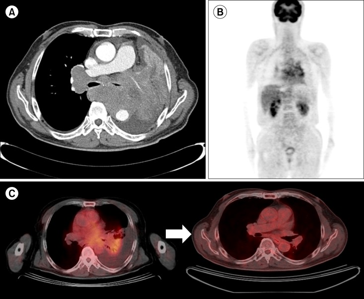
Fig. 3 (A) Chest computed tomography showing conglomerated enlarged lymph nodes in the bilateral mediastinum causing compression of the central airway, and encasement of the left pulmonary artery and descending aorta. (B) A positron emission tomography (PET) image showing a hypermetabolic central lung mass in the left lung with direct invasion of the mediastinum and post-obstructive atelectasis. (C) PET image showing decreased extent and activity of hypermetabolic lesions after rituximab, cyclophosphamide, doxorubicin, vincristine, and prednisolone (R-CHOP) chemotherapy.
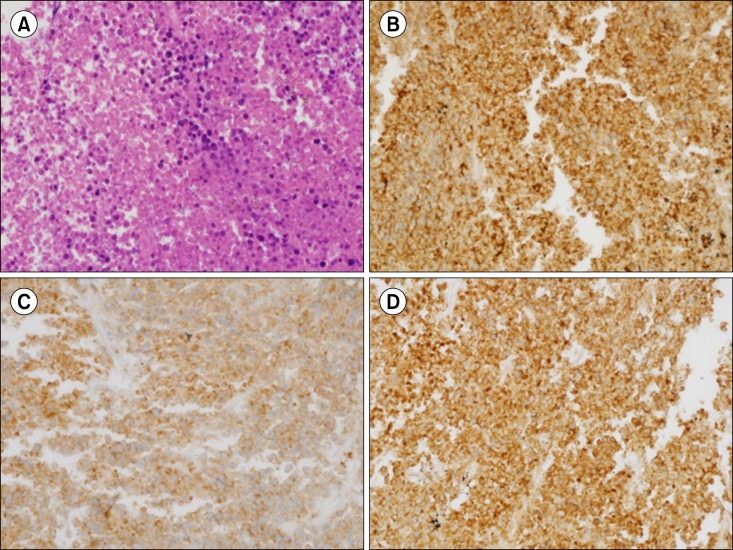
Fig. 4 (A) High magnification shows diffuse infiltration of monotonous tumor cells with a moderate amount of cytoplasm, round-to-oval shaped indented nuclei, condensed chromatin and inconspicuous nucleoli (lymphoblast), and nuclear debris with necrosis (×400; hematoxylin and eosin). (B) The neoplastic cells show diffuse membranous staining for CD20, (C) cytoplasmic staining for CD10, and (D) diffuse membranous staining for leukocyte common antigen (LCA).
Reference
-
1. The Non-Hodgkin's Lymphoma Classification Project. A clinical evaluation of the International Lymphoma Study Group classification of non-Hodgkin's lymphoma. Blood. 1997; 89:3909–3918. PMID: 9166827.2. Pui CH, Behm FG, Crist WM. Clinical and biologic relevance of immunologic marker studies in childhood acute lymphoblastic leukemia. Blood. 1993; 82:343–362. PMID: 8329694.
Article3. Soslow RA, Baergen RN, Warnke RA. B-lineage lymphoblastic lymphoma is a clinicopathologic entity distinct from other histologically similar aggressive lymphomas with blastic morphology. Cancer. 1999; 85:2648–2654. PMID: 10375114.
Article4. Chang MH, Kim SJ, Kim K, et al. Clinical features and treatment outcomes of adult B- and T-lymphoblastic lymphoma: results of multicentre analysis in Korea. Leuk Lymphoma. 2009; 50:1119–1125. PMID: 19557632.
Article5. Lin P, Jones D, Dorfman DM, Medeiros LJ. Precursor B-cell lymphoblastic lymphoma: a predominantly extranodal tumor with low propensity for leukemic involvement. Am J Surg Pathol. 2000; 24:1480–1490. PMID: 11075849.6. Kaygusuz I, Toptas T, Guven A, Firatli-Tuglular T, Tecimer T, Bayik M. Precursor B cell lymphoblastic lymphoma presenting as a solitary bone tumor: a case report and review of the literature. Int J Hematol. 2010; 92:757–761. PMID: 21080126.
Article7. Maitra A, McKenna RW, Weinberg AG, Schneider NR, Kroft SH. Precursor B-cell lymphoblastic lymphoma. A study of nine cases lacking blood and bone marrow involvement and review of the literature. Am J Clin Pathol. 2001; 115:868–875. PMID: 11392884.8. Linker CA, Levitt LJ, O'Donnell M, Forman SJ, Ries CA. Treatment of adult acute lymphoblastic leukemia with intensive cyclical chemotherapy: a follow-up report. Blood. 1991; 78:2814–2822. PMID: 1835410.
Article9. Cortelazzo S, Ponzoni M, Ferreri AJ, Hoelzer D. Lymphoblastic lymphoma. Crit Rev Oncol Hematol. 2011; 79:330–343. PMID: 21273093.
Article10. Soslow RA, Bhargava V, Warnke RA. IC2, TdT, bcl-2, and CD34 expression in paraffin-embedded high-grade lymphoma/acute lymphoblastic leukemia distinguishes between distinct clinicopathologic entities. Hum Pathol. 1997; 28:1158–1165. PMID: 9343323.11. Yoon DH, Sohn BS, Lee WJ, et al. VPDL chemotherapy for T-cell lymphoblastic lymphoma (T-LBL) in adults: comparison with upfront autologous stem cell transplantation in a single center. Korean J Hematol. 2008; 43:138–144.
Article12. Le Gouill S, Lepretre S, Briere J, et al. Adult lymphoblastic lymphoma: a retrospective analysis of 92 patients under 61 years included in the LNH87/93 trials. Leukemia. 2003; 17:2220–2224. PMID: 14576732.
Article13. Reiter A, Schrappe M, Ludwig WD, et al. Intensive ALL-type therapy without local radiotherapy provides a 90% event-free survival for children with T-cell lymphoblastic lymphoma: a BFM group report. Blood. 2000; 95:416–421. PMID: 10627444.
- Full Text Links
-
- Actions
-
Cited
- CITED
-
- Close
- Share
-
- Similar articles
-
- A Case of Non-T,Non-B Primary Cutaneous Lymphoblastic Lymphoma
- Precursor B-Cell Acute Lymphoblastic Leukemia in Two Patients with a History of Cytotoxic Therapy
- Rapid Progression of Mediastinal Tumor within a Few Days: A Case Report of T Cell Lymphoblastic Lymphoma
- Solitary Lymphoblastic Lymphoma of the Thoracic Spine
- Mixed-phenotypic acute leukemia: cytochemically myeloid and phenotypically early T-cell precursor acute lymphoblastic leukemia
