Rapid Progression of Mediastinal Tumor within a Few Days: A Case Report of T Cell Lymphoblastic Lymphoma
- Affiliations
-
- 1Department of Radiology, Seoul Medical Center, Seoul, Korea. ykradio@medimail.co.kr
- 2Department of Hematooncology, Seoul Medical Center, Seoul, Korea.
- 3Department of Pathology, Seoul Medical Center, Seoul, Korea.
- 4Department of Thoracic Surgery, Seoul Medical Center, Seoul, Korea.
- KMID: 2164826
- DOI: http://doi.org/10.3348/jksr.2016.74.5.308
Abstract
- T-cell lymphoblastic lymphoma is a highly aggressive tumor derived from lymphocyte of the thymus, which accounts for 2% of non-Hodgkin's lymphoma. The disease occurs most commonly in adolescent and young adult males. It often results in respiratory emergency because of high proliferation rate. In this case, we confirmed the rapid progression of T-cell lymphoblastic lymphoma through the chest CT scan with one week interval. Three days of empirical chemotherapy resulted in substantial reduction of mediastinal mass, pleural thickening and pleural effusion.
MeSH Terms
Figure
-
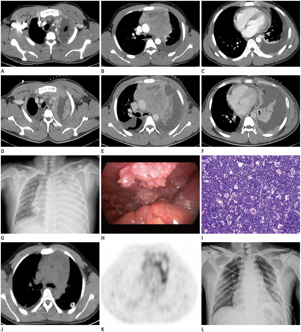
Fig. 1 A 26-year-old man with mediastinal T-cell lymphoblastic lymphoma. A-C. Initial contrast enhanced CT images show a large lobulated anterior mediastinal mass with mass effect, as well as left pleural effusion with nodular thickening. D-F. Follow-up CT scan obtained 7 days after the initial CT shows interval increased size of anterior mediastinal mass and markedly increased amount of left pleural effusion with nodular thickening. G. Chest PA obtained at 7 days after the initial CT shows superior mediastinal widening and markedly increased amount of left pleural effusion with increased extent of left upper pleural thickening. Furthermore, tracheal deviation is worsened. H. Video-assisted thoracoscopy shows highly vascularized anterior mediastinal mass and multiple nodular lesions at parietal pleura. I. Photomicrography of histopathological specimen from the anterior mediastinal lesion reveals diffuse infiltration of small to medium-sized lymphoblasts, which have round to oval and irregular shaped nuclei with several mitotic figures (hematoxylin and eosin staining, × 200). In the immunohistochemical staining (not shown), the neoplastic cells show strong cytoplasmic staining for CD3 and TdT, consistent with T lymphoblastic lymphoma. TdT = terminal deoxynucleotidyl transferase J, K. F-18 FDG PET-CT scan shows heterogeneously hypermetabolic mass in the anterior mediastinum (SUVmax = 4.4) and lower pole of left kidney (SUVmax measured 4.4). L. Chest PA radiography at 7 days after surgical biopsy shows decreased size of the anterior mediastinal mass. The trachea is in normal position. FDG PET-CT = fluorodeoxyglucose positron emission tomography-computed tomography, SUVmax = maximum standardized uptake value
Reference
-
1. Han X, Kilfoy B, Zheng T, Holford TR, Zhu C, Zhu Y, et al. Lymphoma survival patterns by WHO subtype in the United States, 1973-2003. Cancer Causes Control. 2008; 19:841–858.2. Hoelzer D, Gökbuget N. T-cell lymphoblastic lymphoma and T-cell acute lymphoblastic leukemia: a separate entity? Clin Lymphoma Myeloma. 2009; 9:Suppl 3. S214–S221.3. Thomas DA, Kantarjian HM. Lymphoblastic lymphoma. Hematol Oncol Clin North Am. 2001; 15:51–95. vi4. Sweetenham JW. Highly aggressive lymphomas in adults. Hematol Oncol Clin North Am. 2008; 22:965–978. ix5. Willemssen F, Colla R, Vandevenne JE, Palmers Y. Mediastinal T-cell lymphoblastic lymphoma. JBR-BTR. 2002; 85:172–173.6. Tateishi U, Müller NL, Johkoh T, Onishi Y, Arai Y, Satake M, et al. Primary mediastinal lymphoma: characteristic features of the various histological subtypes on CT. J Comput Assist Tomogr. 2004; 28:782–789.
