Changes of Musculoskeletal Deformity in Severely Disabled Children Using the Custom Molded Fitting Chair
- Affiliations
-
- 1Department of Physical & Rehabilitation Medicine, Inha University School of Medicine, Incheon, Korea. recolo2009@gmail.com
- 2Technical Aid Center, Notre Dame Welfare Center, Incheon, Korea.
- KMID: 2266648
- DOI: http://doi.org/10.5535/arm.2013.37.1.33
Abstract
OBJECTIVE
To know the effectiveness of a custom molded fitting chair between pre- and post-chair status through comparison of musculoskeletal indices in severely disabled children.
METHODS
We researched 34 severely disabled patients who had used a custom molded fitting chair continuously for more than a year. There were 27 cerebral palsy patients and 7 patients with other kinds of diseases that affect the brain such as chromosomal disease or metabolic disease. By radiographic studies, Cobb's angle, the femoral neck-shaft angle of the femur, and Reimers migration percentage were measured. The indices are analyzed before and after application.
RESULTS
The average period of application was 24 months. There was a significant reduction in the angles of femur neck-shaft, 163.4 degree before and 158.2 degree after the use of the chair (p<0.05), and 23 of 34 had demonstrated a reduced angle. Cobb's angle and Reimers migration percentage increased but the difference of pre- and post-chair status was not statistically significant. Seventeen of 33 children showed reduced Cobb's angle. Also, 19 of 37 showed a reduced degree of dislocation of the hip joints.
CONCLUSION
In spite of the use of a custom molded fitting chair, a significant improvement did not emerge for musculoskeletal deformity indices in severely disabled children. However, there was no significant aggravation of Cobb's angle or Reimers migration percentage in developing children. Therefore, it is thought be helpful to prevent rapid aggravation of musculoskeletal deformities.
MeSH Terms
Figure
-
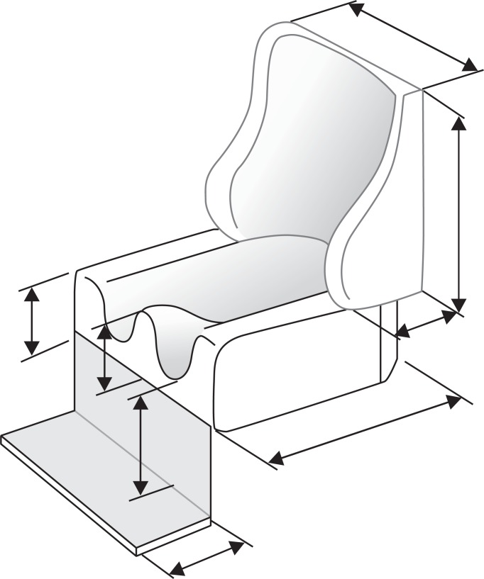
Fig. 1 The blueprint making an inner for the chair.
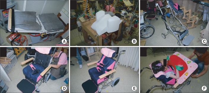
Fig. 2 Manufacturing process of the custom molded fitting chair. Simulator scanned the body shape of the patient (A). The inner was reconstructed from the image scanned by the simulator (B). A frame of a manual wheelchair was selected to apply the inner (C). The seat portion of the inner was installed to the frame. A fan is attached under the back for ventilation (D). The back portion was installed (E). Applying the patient to the chair, a few adjustments were needed (F).
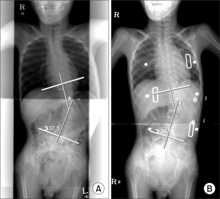
Fig. 3 Cobb's angle (*) measurement in scoliosis. (A) Before application of the chair. (B) After application of the chair.
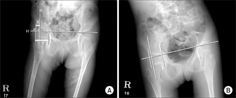
Fig. 4 Reimers migration percentage in hip subluxation. Perkins' line (P), which is drawn perpendicular to Hilgenreiner's line (H). Reimers migration percentage is the percentage of the femoral head lying lateral to Perkins' line (P). Reimers migration percentage=X/Y×100. (A) Before application of the chair. (B) After application of the chair.
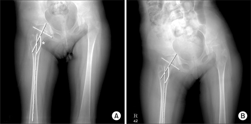
Fig. 5 Femoral neck-shaft (*) angle in coxa valga. (A) Before application of the chair. (B) After application of the chair.
Reference
-
1. Morrell DS, Pearson JM, Sauser DD. Progressive bone and joint abnormalities of the spine and lower extremities in cerebral palsy. Radiographics. 2002; 22:257–268. PMID: 11896216.
Article2. Thometz JG, Simon SR. Progression of scoliosis after skeletal maturity in institutionalized adults who have cerebral palsy. J Bone Joint Surg Am. 1988; 70:1290–1296. PMID: 3182881.
Article3. Madigan RR, Wallace SL. Scoliosis in the institutionalized cerebral palsy population. Spine (Phila Pa 1976). 1981; 6:583–590. PMID: 7336281.
Article4. Miller F, Bagg MR. Age and migration percentage as risk factors for progression in spastic hip disease. Dev Med Child Neurol. 1995; 37:449–455. PMID: 7768344.5. Saito N, Ebara S, Ohotsuka K, Kumeta H, Takaoka K. Natural history of scoliosis in spastic cerebral palsy. Lancet. 1998; 351:1687–1692. PMID: 9734885.
Article6. Gajdosik CG, Cicirello N. Secondary conditions of the musculoskeletal system in adolescents and adults with cerebral palsy. Phys Occup Ther Pediatr. 2001; 21:49–68. PMID: 12043172.
Article7. Greiner KA. Adolescent idiopathic scoliosis: radiologic decision-making. Am Fam Physician. 2002; 65:1817–1822. PMID: 12018804.8. Reimers J. The stability of the hip in children: a radiological study of the results of muscle surgery in cerebral palsy. Acta Orthop Scand Suppl. 1980; 184:1–100. PMID: 6930145.
Article9. Noble PC, Alexander JW, Lindahl LJ, Yew DT, Granberry WM, Tullos HS. The anatomic basis of femoral component design. Clin Orthop Relat Res. 1988; (235):148–165. PMID: 3416522.
Article10. Suh YJ, Kim ES, Park ES, Park HS, Yoon YK, Cho SR. Attenuation of spinal curvature and pelvic obliquity by body shape molded inner system in cerebral palsy with non-fixed scoliosis. J Korean Acad Rehabil Med. 2011; 35:259–264.11. Hoffer MM. Management of the hip in cerebral palsy. J Bone Joint Surg Am. 1986; 68:629–631. PMID: 3514626.
Article12. Ferguson RL, Allen BL Jr. Considerations in the treatment of cerebral palsy patients with spinal deformities. Orthop Clin North Am. 1988; 19:419–425. PMID: 3357689.
Article13. Foroohar A, McCarthy JJ, Yucha D, Clarke S, Brey J. Head-shaft angle measurement in children with cerebral palsy. J Pediatr Orthop. 2009; 29:248–250. PMID: 19305274.
Article
- Full Text Links
-
- Actions
-
Cited
- CITED
-
- Close
- Share
-
- Similar articles
-
- Effect of Custom-Molded Foot Orthoses on Foot Pain and Balance in Children With Symptomatic Flexible Flat Feet
- Effect of Foot Orthoses on Children With Lower Extremity Growing Pains
- Increase of Independence in a Hemipelvectomy Patient with a Custom-Molded Supportive Seating and a Cosmetic Prosthesis : A case report
- Long Term Effect of Custom-Molded Foot Orthoses on Foot Pain and Balance in Children with Symptomatic Flexible Flat Feet
- A Survey of the Outpatient General Anesthesia and Dental Treatment in Chungnam Dental Clinic for the Disabled
