Ann Rehabil Med.
2013 Oct;37(5):683-689. 10.5535/arm.2013.37.5.683.
Ultrasonographic Evaluation of Therapeutic Effects of Complex Decongestive Therapy in Breast Cancer-Related Lymphedema
- Affiliations
-
- 1Department of Physical Medicine and Rehabilitation, Kosin University College of Medicine, Busan, Korea. oggum@daum.net
- KMID: 2266586
- DOI: http://doi.org/10.5535/arm.2013.37.5.683
Abstract
OBJECTIVE
To evaluate the usefulness of ultrasonography as a follow-up tool for evaluating the effects of complex decongestive physiotherapy (CDPT) in breast cancer-related lymphedema (BCRL).
METHODS
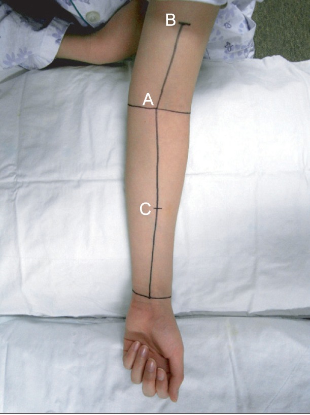
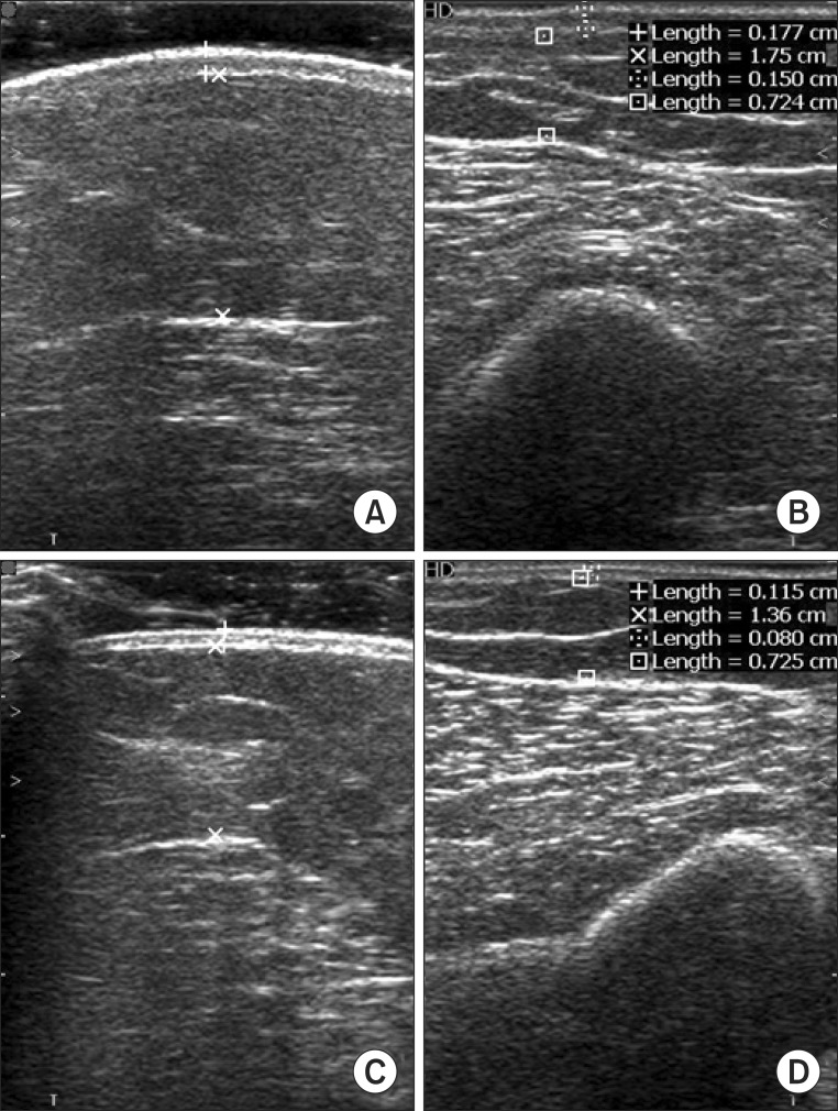
Twenty patients with BCRL were enrolled in this study. All patients had undergone therapy in the CDPT program for 2 weeks. Soft tissue thickness of both the affected and unaffected upper limb was measured before and after CDPT. The measurements were taken at 3 points (the mid-point between the medial and lateral epicondyles at the elbow level, 10 cm proximal and 10 cm distal to the elbow) with and without pressure. We then calculated the compliance of soft tissue before and after CDPT. Circumferences of both the affected and unaffected upper limb were also measured before and after CDPT at the 3 defined points.
RESULTS
After 2 weeks of the CDPT program, the circumference and soft tissue thickness of the unaffected upper limb did not significantly change. In the affected upper limb, the circumference was significantly reduced in the 3 point, when compared with measurements taken prior to treatment. Additionally, soft tissue thickness was significantly reduced at the elbow and 10 cm proximal to the elbow. After CDPT, compliance at each of the 3 points had increased, but this trend was not significantly different.
CONCLUSION
Our results showed that arm circumference and ultrasonography-derived soft tissue thickness was useful as a way of assessing therapeutic effects of CDPT.
MeSH Terms
Figure
Reference
-
1. Jeong HJ, Sim YJ, Hwang KH, Kim GC. Cause of shoulder pain in women with breast cancer-related lymphedema: a pilot study. Yonsei Med J. 2011; 52:661–667. PMID: 21623610.2. Han NM, Cho YJ, Hwang JS, Kim HD, Cho GY. Usefulness of ultrasound examination in evaluation of breast cancer-related lymphedema. J Korean Acad Rehabil Med. 2011; 35:101–109.3. Petrek JA, Pressman PI, Smith RA. Lymphedema: current issues in research and management. CA Cancer J Clin. 2000; 50:292–307. PMID: 11075239.
Article4. Hwang JH, Choi JY, Lee JY, Hyun SH, Choi Y, Choe YS, et al. Lymphscintigraphy predicts response to complex physical therapy in patients with early stage extremity lymphedema. Lymphology. 2007; 40:172–176. PMID: 18365531.5. Deltombe T, Jamart J, Recloux S, Legrand C, Vandenbroeck N, Theys S, et al. Reliability and limits of agreement of circumferential, water displacement, and optoelectronic volumetry in the measurement of upper limb lymphedema. Lymphology. 2007; 40:26–34. PMID: 17539462.6. Tomczak H, Nyka W, Lass P. Lymphoedema: lymphoscintigraphy versus other diagnostic techniques: a clinician's point of view. Nucl Med Rev Cent East Eur. 2005; 8:37–43. PMID: 15977145.7. Stanton AW, Badger C, Sitzia J. Non-invasive assessment of the lymphedematous limb. Lymphology. 2000; 33:122–135. PMID: 11019400.8. Lim CY, Seo HG, Kim K, Chung SG, Seo KS. Measurement of lymphedema using ultrasonography with the compression method. Lymphology. 2011; 44:72–81. PMID: 21949976.9. Kim W, Chung SG, Kim TW, Seo KS. Measurement of soft tissue compliance with pressure using ultrasonography. Lymphology. 2008; 41:167–177. PMID: 19306663.10. Hwang JH, Lee KW, Kwon JY, Kim BT, Choi JY, Lee BB, et al. Improvement of lymphatic function after complex physical therapy change of lymphoscintigraphy. J Korean Acad Rehabil Med. 1998; 22:698–704.
- Full Text Links
- Actions
-
Cited
- CITED
-
- Close
- Share
- Similar articles
-
- The effects of complex decongestive therapy on pain and functionality in individuals with breast cancer who developed adhesive capsulitis due to lymphedema: an evaluation by an isokinetic computerized system
- Effects of Extracorporeal Shockwave Therapy on Improvements in Lymphedema, Quality of Life, and Fibrous Tissue in Breast Cancer-Related Lymphedema
- Long-Term Effects of Complex Decongestive Therapy in Breast Cancer Patients With Arm Lymphedema After Axillary Dissection
- The Effectiveness of Complex Decongestive Physical Therapy for Pain in Patients with Rheumatoid Lymphedema: Two Case Reports
- Feasibility of Extracorporeal Shock Wave Therapy for Complex Upper Limb Morbidity in Breast Cancer Patient


