Congenital Muscular Torticollis Concurrent With Sagittal Synostosis: A Case Report
- Affiliations
-
- 1The Clinic for Torticollis, Department of Physical Medicine and Rehabilitation, Ajou University School of Medicine, Suwon, Korea. syyim@ajou.ac.kr
- KMID: 2266509
- DOI: http://doi.org/10.5535/arm.2014.38.5.712
Abstract
- Congenital muscular torticollis (CMT) and craniosynostosis are diseases that cause plagiocephaly and craniofacial asymmetry in children. In our literature review, we did not find any report of concurrent manifestation of CMT and craniosynostosis. A 41-month-old boy visited our hospital with left torticollis, right laterocollis, and craniofacial asymmetry as the main findings. During clinical examination, prominent right sternocleidomastoid muscle and limited range of motion of the neck were noted, and right CMT was confirmed by magnetic resonance imaging of the neck. Three-dimensional computed tomography of the skull, which was conducted due to the unusual appearance of the skull with a large head circumference, mild brachycephaly, as well as left plagiocephaly, revealed premature closure of the sagittal suture. Thus, we report the first case that showed concurrence of CMT and sagittal synostosis. We recommend that concurrently manifested craniosynostosis needs to be examined if the subject with CMT displays unusual craniofacial asymmetry to a greater extent than deformational plagiocephaly.
Keyword
MeSH Terms
Figure
-
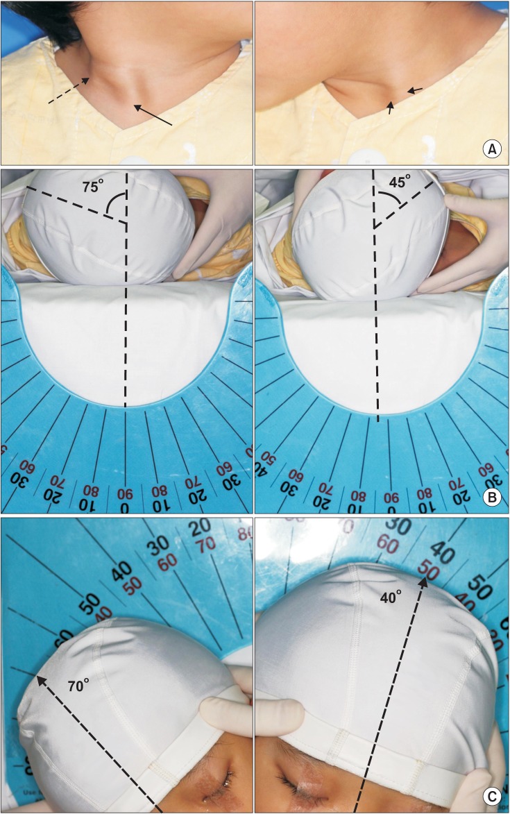
Fig. 1 A 41-month-old boy shows (A) sternal (solid arrow) and clavicular (dotted arrow) heads of the right sternocleidomastoid muscle, which are more prominent than those on the left side (short arrows). (B) Measurement of the range of motion for cervical rotation shows a 30° deficit in the cervical rotation to the right side and (C) a 30° deficit in lateral flexion to the left side compared with that on the contralateral side.
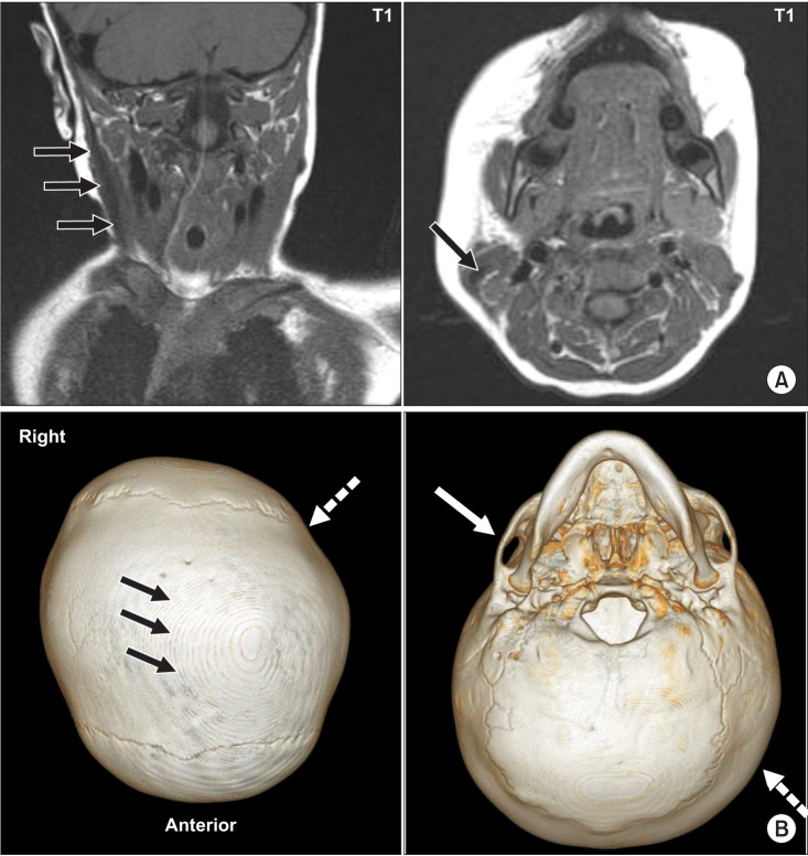
Fig. 2 (A) T1-weighted coronal and axial magnetic resonance images of the neck display fusiform thickening and low signal intensity within the right sternocleidomastoid muscle (solid arrow). (B) Three-dimensional computed tomography of the brain shows premature closure of the sagittal suture (black arrows) with left occipital plagiocephaly (dotted arrow) and flattened right zygomatic arch (white arrow).
Reference
-
1. Looman WS, Flannery AB. Evidence-based care of the child with deformational plagiocephaly, Part I: assessment and diagnosis. J Pediatr Health Care. 2012; 26:242–250. PMID: 22726709.
Article2. Badve CA, K MM, Iyer RS, Ishak GE, Khanna PC. Craniosynostosis: imaging review and primer on computed tomography. Pediatr Radiol. 2013; 43:728–742. PMID: 23636536.
Article3. Bendon CL, Sheerin FB, Wall SA, Johnson D. The relationship between scaphocephaly at the skull vault and skull base in sagittal synostosis. J Craniomaxillofac Surg. 2014; 42:245–249. PMID: 23800755.
Article4. Hwang JH, Lee HB, Kim JH, Park MC, Kwack KS, Han JD, et al. Magnetic resonance imaging as a determinant for surgical release of congenital muscular torticollis: correlation with the histopathologic findings. Ann Rehabil Med. 2012; 36:320–327. PMID: 22837966.
Article5. Raco A, Raimondi AJ, De Ponte FS, Brunelli A, Bristot R, Bottini DJ, et al. Congenital torticollis in association with craniosynostosis. Childs Nerv Syst. 1999; 15:163–169. PMID: 10361966.
Article6. Koljonen V, Leikola J, Valanne L, Hukki J. Atypical craniosynostosis with torticollis and neurological symptoms: a rhombencephalosynapsis sequence. Case Rep Med. 2009; 2009:919463. PMID: 20029674.
Article7. Seo SJ, Yim SY, Lee IJ, Han DH, Kim CS, Lim H, et al. Is craniofacial asymmetry progressive in untreated congenital muscular torticollis? Plast Reconstr Surg. 2013; 132:407–413. PMID: 23584628.
Article8. Lajeunie E, Le Merrer M, Bonaiti-Pellie C, Marchac D, Renier D. Genetic study of scaphocephaly. Am J Med Genet. 1996; 62:282–285. PMID: 8882788.
Article9. Kirschner RE, Gannon FH, Xu J, Wang J, Karmacharya J, Bartlett SP, et al. Craniosynostosis and altered patterns of fetal TGF-beta expression induced by intrauterine constraint. Plast Reconstr Surg. 2002; 109:2338–2354. PMID: 12045561.
- Full Text Links
-
- Actions
-
Cited
- CITED
-
- Close
- Share
-
- Similar articles
-
- Familial Congenital Muscular Torticollis: A Case Report
- Two Cases of Sternomastoid Tumor
- Congenital Torticollis with Bilateral Sternocleidomastoid Muscle Contracture
- Congenital Muscular Torticollis in Siblings: A case report and literature review
- Unipolar Release of the Sternocleidomastoideus in Congenital Muscular Torticollis in Children
