Ann Dermatol.
2013 Feb;25(1):124-126. 10.5021/ad.2013.25.1.124.
Localized Involutional Lipoatrophy in a Child
- Affiliations
-
- 1Department of Dermatology, Sanggye Paik Hospital, Inje University College of Medicine, Seoul, Korea. woods75@hanmail.net
- KMID: 2265991
- DOI: http://doi.org/10.5021/ad.2013.25.1.124
Abstract
- No abstract available.
Figure
-
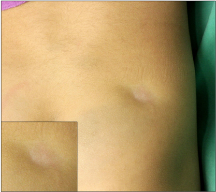
Fig. 1 Well-demarcated, hypopigmented, and depressed atrophic patch on the right buttock.
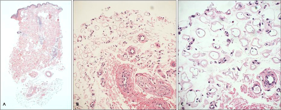
Fig. 2 Histological examination of the skin biopsy specimen shows. (A) Epidermal atrophy, compaction of collagen bundle in the dermis without inflammation (H&E, ×40). (B) Diminutive fat lobules composed of variable sizes adipocytes (H&E, ×200). (C) Immature adipocytes resembling fetal fat tissue (H&E, ×400).
Reference
-
1. Dahl PR, Zalla MJ, Winkelmann RK. Localized involutional lipoatrophy: a clinicopathologic study of 16 patients. J Am Acad Dermatol. 1996. 35:523–528.
Article2. Peters MS, Winkelmann RK. The histopathology of localized lipoatrophy. Br J Dermatol. 1986. 114:27–36.
Article3. Yamamoto T, Yokozeki H, Nishioka K. Localized involutional lipoatrophy: report of six cases. J Dermatol. 2002. 29:638–643.
Article4. Abbas O, Salman S, Kibbi AG, Chedraoui A, Ghosn S. Localized involutional lipoatrophy with epidermal and dermal changes. J Am Acad Dermatol. 2008. 58:490–493.
Article5. Ahmed I. Post-injection involutional lipoatrophy: ultrastructural evidence for an activated macrophage phenotype and macrophage related involution of adipocytes. Am J Dermatopathol. 2006. 28:334–337.
