Induction of a Hardening Phenomenon and Quantitative Changes of Ceramides in Stratum Corneum
- Affiliations
-
- 1Department of Dermatology, Hallym University Kangnam Sacred Heart Hospital, Seoul, Korea. hyeonekim@gmail.com
- KMID: 2265695
- DOI: http://doi.org/10.5021/ad.2014.26.1.35
Abstract
- BACKGROUND
Hardening phenomenon of human skin after repeated exposure to the irritants is well-known, but the precise mechanism remains elusive.
OBJECTIVE
To modify the previous experimental model of hardening phenomenon by repeated applications of two different concentrations of sodium lauryl sulfate (SLS) solutions to Korean healthy volunteers and to investigate the quantitative changes of ceramides in stratum corneum before and after chronic repeated irritation.
METHODS
Eight hundred microliters of distilled water containing 0.1% and 2% SLS was applied for 10 minutes on the forearm of 41 healthy volunteers for 3 weeks. After an intervening 3-week rest, 24-hour patch tests with 1% SLS were conducted on previously irritated sites. Transepidermal water loss (TEWL), erythema index and quantity of ceramide were measured in the stratum corneum before and after irritation.
RESULTS
TEWL values on the sites preirritated with 2% SLS were lower than those with 0.1% SLS. Hardening phenomenon occurred in 24 volunteers at day 44. The changes in ceramide levels were not significantly higher in the hardened skin than in the non-hardened skin.
CONCLUSION
Repetitive stimulation with a higher concentration of SLS can more easily trigger skin hardening.
MeSH Terms
Figure
-
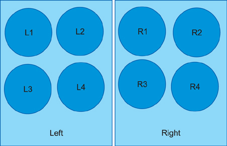
Fig. 1 Pattern of the eight, 20-mm ring marks on the left and right forearms.
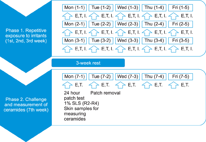
Fig. 2 Schematic illustration of the study design. During the first 3 weeks, repetitive applications of irritants (I sites L1 and R1 for the controls, L2 and R2 for distilled water sites, L3 and R3 for 0.1% sodium lauryl sulfate [SLS], sites L4 and R4 for 2% SLS) on both forearms of the volunteers were performed 5 times per week and erythema index (E) and TEWL (T) were measured before each application (Phase 1). After an intervening 3-weeks, the 24-hour patch test with 1% SLS was performed on sites R2, R3 and R4 and skin samples were obtained from sites L1, L2, L3 and L4. E and T were measured before and after the patch test (7-1 to 7-5) (Phase 2).
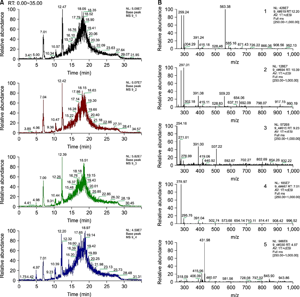
Fig. 3 Examples of high performance liquid chromatography chromatograms of extracted stratum corneum lipid samples of sites L1~L4 sites of one person (A). Examples of tandem mass spectrometry patterns taken from the left fourth chromatogram. One, 2 and 3 show ceramide mass spectrometry pattern (B). RT: retention time, MS: mass spectrometry.
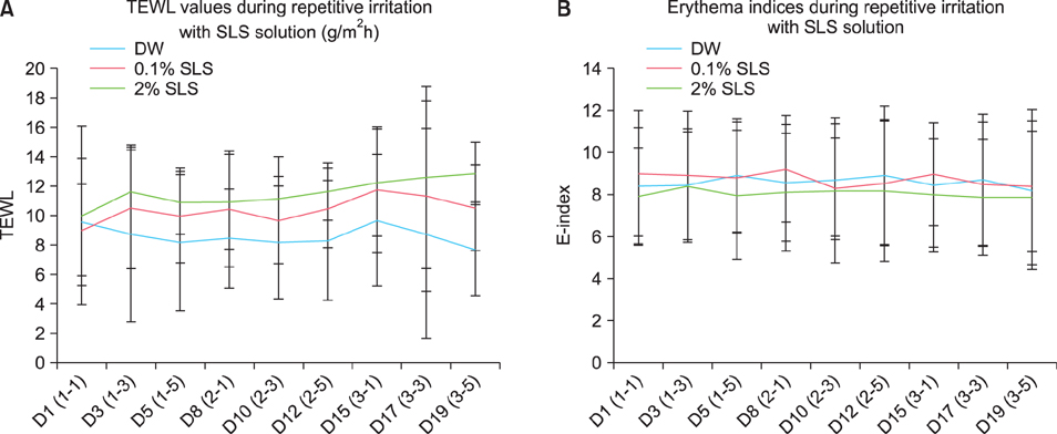
Fig. 4 Transepidermal water loss (TEWL) (g/m2/h) (A) and erythema index (E-index) (B) on the sites stimulated with distilled water (DW), 0.1% sodium lauryl sulfate (SLS) and 2% SLS (Phase 1). The differences in TEWL and E-index between test materials were not statistically significant (p<0.05).
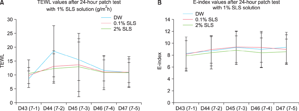
Fig. 5 Transepidermal water loss (TEWL) (g/m2/h) (A) and erythema index (E-index) (B) on the sites exposed to the irritants and those not exposed to the irritants sites after challenging with the 24-hour patch test with 1% sodium lauryl sulfate (SLS) (Phase 2). The 24-hour patch test was performed on days 43 and 44. The increase in TEWL after removing the patch at day 44 was higher on the sites exposed to distilled water (DW) than on those exposed to the two concentrations of SLS (p<0.05). The increase E-index after the 24-hour patch test was lower on the sites exposed to 2% SLS than on those exposed to 0.1% SLS or distilled water. However, the differences were not statistically significant.
Reference
-
1. Smith HR, Basketter DA, McFadden JP. Irritant dermatitis, irritancy and its role in allergic contact dermatitis. Clin Exp Dermatol. 2002; 27:138–146.
Article2. John SM, Uter W, Schwanitz HJ. Relevance of multiparametric skin bioengineering in a prospectively-followed cohort of junior hairdressers. Contact Dermatitis. 2000; 43:161–168.
Article3. van der Valk PG, Nater JP, Bleumink E. Skin irritancy of surfactants as assessed by water vapor loss measurements. J Invest Dermatol. 1984; 82:291–293.
Article4. Agner T. Basal transepidermal water loss, skin thickness, skin blood flow and skin colour in relation to sodiumlauryl-sulphate-induced irritation in normal skin. Contact Dermatitis. 1991; 25:108–114.
Article5. Agner T, Serup J, Handlos V, Batsberg W. Different skin irritation abilities of different qualities of sodium lauryl sulphate. Contact Dermatitis. 1989; 21:184–188.
Article6. Agner T, Serup J. Seasonal variation of skin resistance to irritants. Br J Dermatol. 1989; 121:323–328.
Article7. Watkins SA, Maibach HI. The hardening phenomenon in irritant contact dermatitis: an interpretative update. Contact Dermatitis. 2009; 60:123–130.
Article8. di Nardo A, Sugino K, Wertz P, Ademola J, Maibach HI. Sodium lauryl sulfate (SLS) induced irritant contact dermatitis: a correlation study between ceramides and in vivo parameters of irritation. Contact Dermatitis. 1996; 35:86–91.
Article9. Slodownik D, Lee A, Nixon R. Irritant contact dermatitis: a review. Australas J Dermatol. 2008; 49:1–9.
Article10. Heinemann C, Paschold C, Fluhr J, Wigger-Alberti W, Schliemann-Willers S, Farwanah H, et al. Induction of a hardening phenomenon by repeated application of SLS: analysis of lipid changes in the stratum corneum. Acta Derm Venereol. 2005; 85:290–295.
Article11. de Jongh CM, Lutter R, Verberk MM, Kezic S. Differential cytokine expression in skin after single and repeated irritation by sodium lauryl sulphate. Exp Dermatol. 2007; 16:1032–1040.
Article12. Branco N, Lee I, Zhai H, Maibach HI. Long-term repetitive sodium lauryl sulfate-induced irritation of the skin: an in vivo study. Contact Dermatitis. 2005; 53:278–284.
Article13. Widmer J, Elsner P, Burg G. Skin irritant reactivity following experimental cumulative irritant contact dermatitis. Contact Dermatitis. 1994; 30:35–39.
Article14. Robinson MK. Intra-individual variations in acute and cumulative skin irritation responses. Contact Dermatitis. 2001; 45:75–83.
Article15. Zettersten EM, Ghadially R, Feingold KR, Crumrine D, Elias PM. Optimal ratios of topical stratum corneum lipids improve barrier recovery in chronologically aged skin. J Am Acad Dermatol. 1997; 37:403–408.
Article16. van der Valk PG, Maibach HI. A functional study of the skin barrier to evaporative water loss by means of repeated cellophane-tape stripping. Clin Exp Dermatol. 1990; 15:180–182.
Article17. McOsker DE, Beck LW. Characteristics of accommodated (hardened) skin. J Invest Dermatol. 1967; 48:372–383.18. Bouwstra JA, Gooris GS, Dubbelaar FE, Weerheim AM, Ijzerman AP, Ponec M. Role of ceramide 1 in the molecular organization of the stratum corneum lipids. J Lipid Res. 1998; 39:186–196.
Article19. Proksch E. The epidermis as metabolically active tissue: regulation of lipid synthesis by the barrier function. Z Hautkr. 1990; 65:296–300.20. Imokawa G, Abe A, Jin K, Higaki Y, Kawashima M, Hidano A. Decreased level of ceramides in stratum corneum of atopic dermatitis: an etiologic factor in atopic dry skin. J Invest Dermatol. 1991; 96:523–526.
Article21. Yamamoto A, Serizawa S, Ito M, Sato Y. Stratum corneum lipid abnormalities in atopic dermatitis. Arch Dermatol Res. 1991; 283:219–223.
Article22. de Jongh CM, Khrenova L, Verberk MM, Calkoen F, van Dijk FJ, Voss H, et al. Loss-of-function polymorphisms in the filaggrin gene are associated with an increased susceptibility to chronic irritant contact dermatitis: a case-control study. Br J Dermatol. 2008; 159:621–627.
Article
- Full Text Links
-
- Actions
-
Cited
- CITED
-
- Close
- Share
-
- Similar articles
-
- Epidermal Lipid Homeostasis
- Effect of Delipidization on Binding of Hydrocortisone to Human Stratum Corneum
- A Morphological Study of Stratum Corneum
- The Ultrastructural Changes of Stratum Corneum Lipids after Application of Oleic Acid in Propylene Glycol
- The Assessment of the Efficiency of Lipid Extraction by Several Solvents from the Stratum Corneum
