Ann Dermatol.
2013 Nov;25(4):520-522. 10.5021/ad.2013.25.4.520.
Recurrent Milia-Like Idiopathic Calcinosis Cutis on the Upper Eyelid
- Affiliations
-
- 1Department of Dermatology, Chosun University School of Medicine, Gwangju, Korea. kimminsung@chosun.ac.kr
- KMID: 2265082
- DOI: http://doi.org/10.5021/ad.2013.25.4.520
Abstract
- No abstract available.
MeSH Terms
Figure
-

Fig. 1 (A) Several milia-like whitish papules on the right upper eyelid (6 years ago). (B) A solitary, firm, 5 mm sized whitish papule was observed in the same area (at present). The authors are indebted to the patient for his permission for publication.
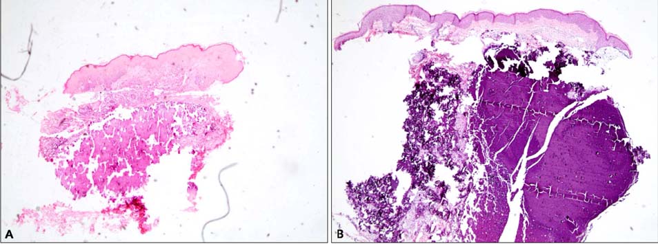
Fig. 2 (A) Amorphous basophilic material surrounded by collagen fibers and fibroblasts in the papillary dermis (H&E, ×40). (B) Amorphous homogenous and basophilic material that stained black with Von Kossa stain in the upper dermis (H&E, ×40).
Reference
-
1. Kim DH, Kang H, Cho SH, Park YM. Solitary milialike idiopathic calcinosis cutis unassociated with Down's syndrome: two case reports. Acta Derm Venereol. 2000; 80:151–152.2. Bécuwe C, Roth B, Villedieu MH, Chouvet B, Kanitakis J, Claudy A. Milia-like idiopathic calcinosis cutis. Pediatr Dermatol. 2004; 21:483–485.
Article3. Jang EJ, Lee JY, Yoon TY. Milia-like idiopathic calcinosis cutis occurring in a toddler born as a premature baby. Ann Dermatol. 2011; 23:490–492.
Article4. Lee DW, Yoon DH, Lee YS, Shim SI, Cho BK. Solitary milialike idiopathic calcinosis cutis: a case unassociated with Down syndrome. J Dermatol. 1996; 23:53–55.
- Full Text Links
-
- Actions
-
Cited
- CITED
-
- Close
- Share
-
- Similar articles
-
- A Case of Milia-like Idiopathic Calcinosis Cutis in an Elderly Person
- A Case of Milia-like Idiopathic Calcinosis Cutis in Healthy Boy
- A Case of Milia-like Idiopathic Calcinosis Cutis That Improved with Tretinoin Cream
- Two Cases of Milia-like Idiopathic Calcinosis Cutis Occurred in Infants
- A Case of Milia-like Idiopathic Calcinosis Cutis and Periorbital Syringomas in Down Syndrome
