Anat Cell Biol.
2013 Jun;46(2):167-170. 10.5115/acb.2013.46.2.167.
Right-sided aortic arch with the retroesophageal left subclavian artery as the fourth branch
- Affiliations
-
- 1Department of Anatomy, Chonbuk National University Medical School and Institute for Medical Sciences, Chonbuk National University, Jeonju, Korea. asch@jbnu.ac.kr
- KMID: 2263112
- DOI: http://doi.org/10.5115/acb.2013.46.2.167
Abstract
- We present a rare variation of the right-sided aortic arch with the retroesophageal left subclavian artery as the forth branch found in a cadaver of an 89-year-old Korean woman during a routine dissection. In this case, the first branch that arose from the ascending aorta was the left common carotid artery, which crossed ventral to the trachea in a left cephalic direction, followed by the right common carotid artery and then the right subclavian artery. Distal to these branches the aortic arch ran dorsally, passing between the esophagus and the vertebra. The left subclavian artery arose from the descending portion of the aortic arch, crossing over to the left upper extremity behind the esophagus. This anomaly was not accompanied by congenital heart disease. Accurate information regarding this variation is of great importance to surgeons for its early identification and preservation during interventions and to radiologists for precise interpretation of angiograms.
MeSH Terms
Figure
-
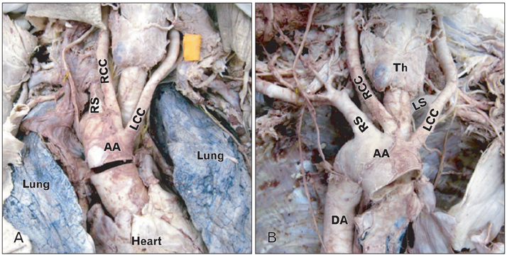
Fig. 1 A ventral view of the right-sided aortic arch before (A) and after (B) the heart and lungs were removed. AA, aortic arch; DA, descending aorta; LCC, left common carotid artery; LS, left subclavian artery; RCC, right common carotid artery; RS, right subclavian artery; Th, thyroid gland.
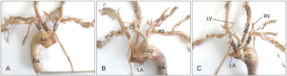
Fig. 2 Photographs of the right-sided aortic arch variation. Ventral (A), medial (B), and dorsal (C) views. AA, aortic arch; AD, aortic diverticulum; DA, descending aorta; LA, ligamentum arteriosum; LCC, left common carotid artery; LS, left subclavian artery; LV, left vertebral artery; RCC, right common carotid artery; RS, right subclavian artery; RV, right vertebral artery.
Reference
-
1. Collins P. Standring S, editor. Development of the cardiovascular and lymphatic system. Gray's Anatomy: The Anatomical Basis of Clinical Practice. 2005. 39th ed. London: Elsevier Churchill Livingstone;1042–1050.2. Moore KL, Persaud TVN. The developing human: clinically oriented embryology. 2003. 8th ed. Philadelpia: Saunders Elsevier.3. Nayak SR, Pai MM, Prabhu LV, D'Costa S, Shetty P. Anatomical organization of aortic arch variations in the India: embryological basis and review. J Vasc Bras. 2006. 5:95–100.4. Alsaif HA, Ramadan WS. An anatomical study of the aortic arch variations. JKAU Med Sci. 2010. 17:37–54.5. Hastreiter AR, D'Cruz IA, Cantez T, Namin EP, Licata R. Right-sided aorta. I. Occurrence of right aortic arch in various types of congenital heart disease. II. Right aortic arch, right descending aorta, and associated anomalies. Br Heart J. 1966. 28:722–739.6. Predey TA, McDonald V, Demos TC, Moncada R. CT of congenital anomalies of the aortic arch. Semin Roentgenol. 1989. 24:96–113.7. Edwards JE. Anomalies of the derivatives of the aortic arch system. Med Clin North Am. 1948. 32:925–949.8. Kommerell B. Verlagerung des Ösophagus durch eine abnorm verlaufende Arteria subclavia dextra (Arteria lusoria). Fortschr Geb Roentgenstrahlen. 1936. 54:590–595.9. Adovasio DA. Congenital anomalies of the aortic arch. Archivio Chir Torace. 1953. 10:171–189.10. Türkvatan A, Büyükbayraktar FG, Olçer T, Cumhur T. Congenital anomalies of the aortic arch: evaluation with the use of multidetector computed tomography. Korean J Radiol. 2009. 10:176–184.
- Full Text Links
-
- Actions
-
Cited
- CITED
-
- Close
- Share
-
- Similar articles
-
- Evaluation of congenital anomalies of the aortic arch by CT and MRI
- Right Aortic Arch with a Retroesophageal Left Subclavian Artery and an Anomalous Origin of the Pulmonary Artery from the Aorta
- Translocation of the Aortic Arch with Norwood Procedure for Hypoplastic Left Heart Syndrome Variant with Circumflex Retroesophageal Aortic Arch
- Surgical Treatment of Occluded Aberrant Left Subclavian Artery with Right-sided Aortic Arch: A case report
- A Case of Right Sided Aortic Arch Causing Superior Vena Cava Syndrome
