Case report of solitary giant hepatic lymphangioma
- Affiliations
-
- 1Department of Surgery, Gimpo Woori Hospital, Gimpo, Korea.
- 2Department of Radiology, Gimpo Woori Hospital, Gimpo, Korea. corbrain@naver.com
- KMID: 2243018
- DOI: http://doi.org/10.14701/kjhbps.2016.20.2.71
Abstract
- A hepatic lymphangioma is a rare benign neoplasm that is usually associated with systemic lymphangiomatosis. A solitary hepatic lymphangioma is extremely rare. Therefore, we present a rare case of a female patient who underwent right hepatectomy for solitary giant hepatic lymphangioma. A 42-year-old female presented to the emergency department with complaint of severe abdominal pain of the right upper quadrant. Abdominal computed tomography showed an approximately 23×30-cm sized, giant, relatively well-defined, homogenous cystic mass with few septa in the right liver (segments VII and VIII). The preoperative diagnosis was a giant hepatic cystadenoma or cystadenocarcinoma. We performed right hepatectomy. The permanent histopathological report revealed cystic lymphangioma of the liver. Although the prognosis of solitary hepatic lymphangioma after surgical resection is favorable, recurrence has been reported in literature.
Keyword
MeSH Terms
Figure
-
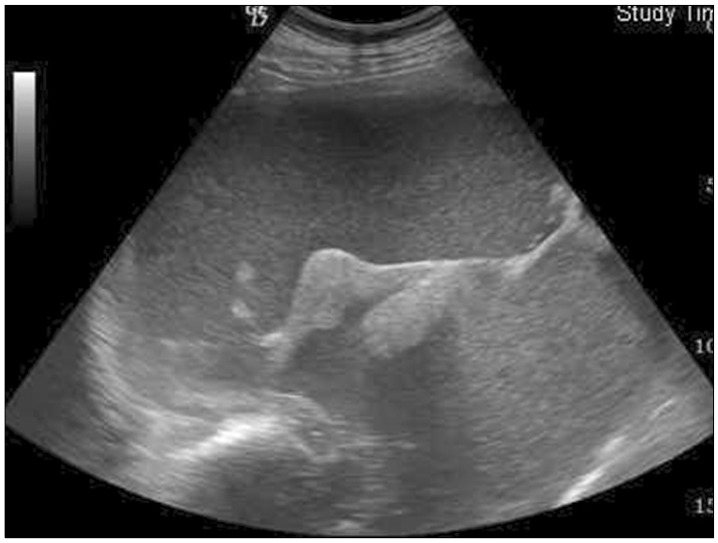
Fig. 1 Abdominal ultrasonography shows a hemorrhagic complicated giant cystic mass with septation and multifocal hyperechoic solid components in the right liver.
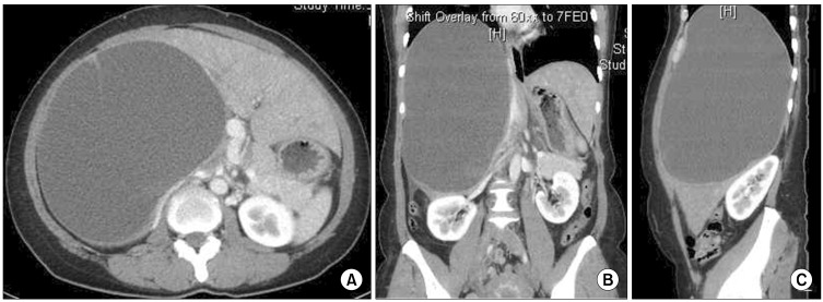
Fig. 2 Abdominal computed tomography images show a giant and relatively well-defined homogenous cystic mass of size 23×30 cm occupying segment VII and VIII of the liver and extending from the diaphragm to the right iliac crest (A-C).
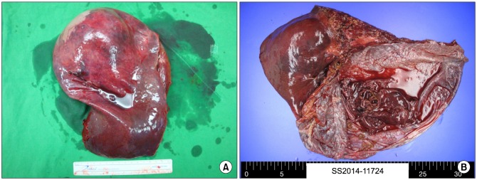
Fig. 3 Gross photographs of the resected specimen (A) and the opened cyst (B) showing a mass of around 23×30 cm in size with a unilocular cyst and pinkish white smooth inner surface, which was filled with coffee-like fluid and sludge.

Fig. 4 Microscopic photographs. Histologically, the cyst wall is lined with single-layered flat endothelial cells (H&E, ×200, A). Immunohistochemical findings revealed these endothelial-lined cells to be positively immunostained by CD31 (×400, B).
Reference
-
1. Stavropoulos M, Vagianos C, Scopa CD, Dragotis C, Androulakis J. Solitary hepatic lymphangioma. A rare benign tumour: a case report. HPB Surg. 1994; 8:33–36. PMID: 7993862.2. Huang L, Li J, Zhou F, Yan J, Liu C, Zhou AY, et al. Giant cystic lymphangioma of the liver. Hepatol Int. 2010; 4:784–787. PMID: 21286352.
Article3. Zhang YZ, Ye YS, Tian L, Li W. Rare case of a solitary huge hepatic cystic lymphangioma. World J Clin Cases. 2013; 1:152–154. PMID: 24303489.
Article4. Liu Q, Sui CJ, Li BS, Gao A, Lu JY, Yang JM. Solitary hepatic lymphangioma: a one-case report. Springerplus. 2014; 3:314. PMID: 25279280.
Article5. Chung JH, Suh YL, Park IA, Jang JJ, Chi JG, Kim YI, et al. A pathologic study of abdominal lymphangiomas. J Korean Med Sci. 1999; 14:257–262. PMID: 10402167.
Article6. Roisman I, Manny J, Fields S, Shiloni E. Intra-abdominal lymphangioma. Br J Surg. 1989; 76:485–489. PMID: 2660949.
Article7. Vargas-Serrano B, Alegre-Bernal N, Cortina-Moreno B, Rodriguez-Romero R, Sanchez-Ortega F. Abdominal cystic lymphangiomas: US and CT findings. Eur J Radiol. 1995; 19:183–187. PMID: 7601168.
Article
