
A Case of Isolated Hepatic Lymphangioma
- Affiliations
-
- 1Department of Internal Medicine, Maryknoll Medical Center, Busan, Korea. clavicle22@hanmail.net
- KMID: 1792826
- DOI: http://doi.org/10.4166/kjg.2012.59.2.189
Abstract
- Hepatic lymphangioma is a rare benign neoplasm. It usually occurs as a part of systemic lymphangiomatosis. Isolated hepatic lymphangioma is extremely rare. A 58-year-old woman with weight loss was referred for the evaluation of chronic renal insufficiency and hepatic mass. Abdominal computed tomography showed 3 cm sized multilobulated cystic lesion with calcification and thick septal enhancing focus in the segment V of the liver. On abdominal magnetic resonance imaging, the masses exhibited low signal intensity on the T1-weighted images and high signal intensity on the T2-weighted images. Malignant tumor could not be ruled out, and therefore, the patient underwent right anterior segmentectomy of the liver. Gross pathology reveraled a 3.0x2.2x1.5 cm mass with multichamber cyst, which was filled with mucoid material. Histologically the mass was composed of irregularly shaped vascular channels filled acellular homogeneous lymph fluids. The final diagnosis was hepatic isolated cavernous lymphangioma. Herein, we report a case of isolated hepatic lymphangioma and also review the existing literature.
Keyword
MeSH Terms
Figure
-
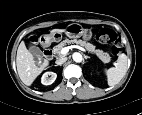
Fig. 1 Abdominal CT finding. It showed 3 cm sized multilobulated cystic lesion with calcification and thick septal enhancing focus in the S5 of the liver.
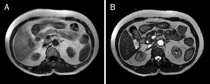
Fig. 2 Abdominal MRI finding. (A) The mass showed low signal intensity on the T1-weighted image and (B) high signal intensity on the T2-weighted image.
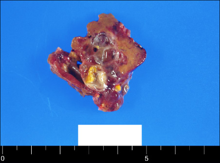
Fig. 3 Gross finding. The cut surface revealed a cyst with multichamber, measuring 3.0×2.2×1.5 cm in dimensions, which was filled with mucoid material.

Fig. 4 Microscopic finding. (A) Mass was composed of irregularly shaped vascular channels filled acellular homogeneous lymph fluids (H&E, ×40). (B) Vascular channels were lined by a single layer of cuboidal cells which reacted positively for factor VIII antigen (peroxidaseanti-peroxidase stain, ×40).
Reference
-
1. Van Steenbergen W, Joosten E, Marchal G, et al. Hepatic lymphangiomatosis. Report of a case and review of the literature. Gastroenterology. 1985. 88:1968–1972.2. Chan SC, Huang SF, Lee WC, Wan YL. Solitary hepatic lymphangioma--a case report. Int J Clin Pract Suppl. 2005. (147):100–102.3. Shahi KS, Geeta B, Rajput P. Solitary hepatic lymphangioma in a 22-day-old infant. J Pediatr Surg. 2009. 44:E9–E11.4. Stavropoulos M, Vagianos C, Scopa CD, Dragotis C, Androulakis J. Solitary hepatic lymphangioma. A rare benign tumour: a case report. HPB Surg. 1994. 8:33–36.5. Yoon DH, Lee JH, Choi DR, et al. A case of isolated hepatic lymphangiomatosis. Korean J Gastroenterol. 2002. 40:348–351.6. Nzegwu MA, Ekenze SO, Okafor OC, Anyanwu PA, Odetunde OA, Olusina DB. Solitary hepatic lymphangioma in an infant. J Perinat Med. 2007. 35:164–165.7. Cutillo DP, Swayne LC, Cucco J, Dougan H. CT and MR imaging in cystic abdominal lymphangiomatosis. J Comput Assist Tomogr. 1989. 13:534–536.8. Wooten WB, Bernardino ME, Goldstein HM. Computed tomography of necrotic hepatic metastases. AJR Am J Roentgenol. 1978. 131:839–842.9. O'Sullivan DA, Torres VE, de Groen PC, Batts KP, King BF, Vockley J. Hepatic lymphangiomatosis mimicking polycystic liver disease. Mayo Clin Proc. 1998. 73:1188–1192.10. Chung JH, Suh YL, Park IA, et al. A pathologic study of abdominal lymphangiomas. J Korean Med Sci. 1999. 14:257–262.11. Kim HH, Hur YH, Park CY, et al. Splenic cavernous lymphangioma mimickimg splenic hemangioma. J Korean Surg Soc. 2009. 77:434–437.12. Ra SH, Bradley RF, Fishbein MC, Busuttil RW, Lu DS, Lassman CR. Recurrent hepatic lymphangiomatosis after orthotopic liver transplantation. Liver Transpl. 2007. 13:1593–1597.13. Datz C, Graziadei IW, Dietze O, et al. Massive progression of diffuse hepatic lymphangiomatosis after liver resection and rapid deterioration after liver transplantation. Am J Gastroenterol. 2001. 96:1278–1281.


