The Role of Neutrophil Estrogen Receptor Status on Maspin Synthesis via Nitric Oxide Production in Human Breast Cancer
- Affiliations
-
- 1Sinha Institute of Medical Science & Technology, Garia, India. asruksinha@yahoo.com
- 2Department of Chemistry, Jadavpur University, Kolkata, India.
- KMID: 2242188
- DOI: http://doi.org/10.4048/jbc.2012.15.2.181
Abstract
- PURPOSE
Estrogen, through its binding to nuclear estrogen receptor (ER), has been implicated in the development of human breast cancer. The presence or absence of ER in breast lesions has been used to classify breast cancer into ER+ or ER- type. Maspin, an anti-breast cancer protein produced in normal mammary cells, has also been reported to control the condition. Studies have been conducted to determine the role of ER+ and ER- status in neutrophils in the synthesis of maspin in human breast cancer.
METHODS
Maspin presence was determined by enzyme linked immunosorbent assay, while nitric oxide (NO) level was determined using the methemoglobin method.
RESULTS
Scatchard plots of the equilibrium binding of estrogen demonstrated the presence of 4.18x10(7) receptors per normal neutrophil and 2.46x10(7) receptors per ER+ neutrophil with a similar dissociation constant (0.926 nM). The ER- type showed nonspecific estrogen binding only. At 0.6 nM estrogen, NO synthesis was maximally increased to 1.829 and 0.887 microM NO/10(9) cells at 4 hours in normal and ER+ neutrophils respectively, with synthesis of 2.383 and 1.422 nM maspin in normal and ER+ neutrophils respectively. Estrogen failed to produce these effects in ER- neutrophils.
CONCLUSION
ER status in neutrophils determined maspin synthesis in breast cancer through the stimulation of NO synthesis. Neutrophils with ER- status which do not produce any maspin when treated with estrogen, might imply a worse prognostic outcome in ER- breast cancer due to the lack of anti-breast cancer protein synthesis.
Keyword
MeSH Terms
Figure
-
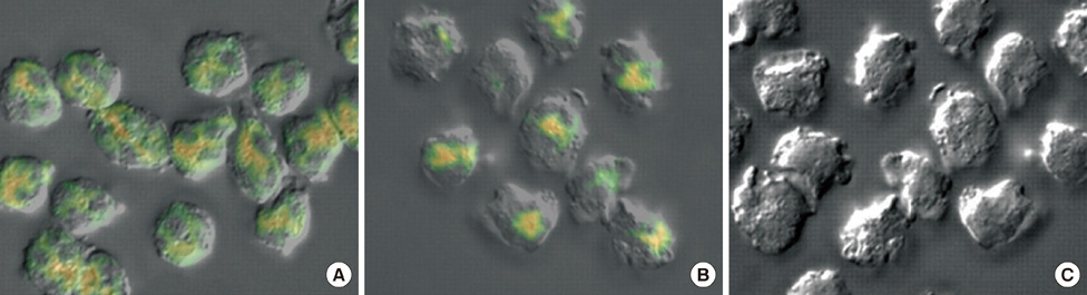
Figure 1 The immunohistochemistry of estrogen receptors (ER) in neutrophils from normal volunteers and patients with breast cancer patients: normal (A), ER+ neutrophils (B), and ER- neutrophils (C). The Figure presented is representative of six or more experiments using neutrophils from six different subjects from each group. The immunohistochemistry of the ER was determined as described in the Methods section. The cells are observed under 45× objective.
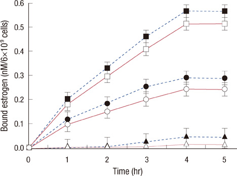
Figure 2 Equilibrium binding of estrogen to normal, estrogen receptor+ (ER+), and ER- neutrophils. Neutrophil suspensions prepared from normal, ER+, and ER- neutrophils were prepared from patients with breast cancer that were treated with 0.6 nM estrogen for different periods of time for equilibrium binding as indicated. The nonspecific binding was determined by adding excess amounts of estrogen (10 mM) to binding mixture containing 1.0 µCi of 14C estradiol as described in the Methods section. The levels estrogen bound to the neutrophils were determined by enzyme linked immunosorbent assay as described in the Methods section. Specific binding was calculated by subtracting the nonspecific binding from the total binding. The solid squares (■) and hollow squares (□) represent total binding and specific binding in normal neutrophils, respectively. The solid circles (●) and hollow circles (◯) indicate total and specific binding in ER+ neutrophils, respectively. The solid triangles (▲) and hollow triangles (△) represent the total and specific binding in neutrophils isolated from ER- subjects, respectively. The solid lines () represent specific binding, while the dotted lines () represent total binding of estrogen to the neutrophils. The results are the mean±SD of five different experiments each in triplicate using blood of five different patients with ER+ or ER- breast cancer or from normal volunteers.
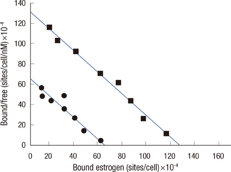
Figure 3 Scatchard plots of the binding of estrogen to neutrophils of normal volunteers, and or patients with estrogen receptor+ (ER+) breast cancer. The neutrophils preparations were suspended in Hank's balanced salt solution (pH 7.4) containing different concentrations of estrogen for 4 hours as indicated for equilibrium binding. The total binding for each point was calculated from the total amount of the ligand present in the reaction mixture. The Scatchard plots shown here are representative of three experiments conducted for each group (n=5). The solid squares (■) represent a Scatchard plot of neutrophils from normal volunteers, while the solid circles (●) indicate a Scatchard plot using neutrophils from patients with ER+ breast cancer.
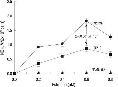
Figure 4 Effect of different amounts of estrogen on the synthesis of nitric oxide (NO) in normal, estrogen receptor (ER)+, or ER- neutrophils. The neutrophil suspensions were prepared from the blood of patients with ER+ or ER- breast cancer and from age-matched normal female volunteers as described in the Methods section (n=15, in each group). The neutrophils preparations (6×10⁹ cells/L) were suspended in Hank's balanced salt solution (pH 7.4) and treated with different concentrations of estrogen (nM) as indicated. In a parallel set of experiments, neutrophil suspensions from age-matched normal female volunteers and from patients with ER+ or ER- breast cancer were incubated with NAME along with different amounts of estrogen. After incubation for 4 hours at 37℃ under sterile conditions, NO synthesis in the reaction mixture was determined using a methemoglobin assay. Solid circles (●) represent NO synthesis in normal neutrophils while, solid squares (■) indicate NO synthesis in ER+ breast cancer neutrophils, the solid triangles (▲) represent NO synthesis in ER- breast cancer neutrophils, and the hollow circles (◯) indicate NO synthesis in presence of NAME. Results are mean±SD of five different experiments in triplicate using blood of 15 different patients with breast cancer in each group and 15 normal female volunteers. NAME=L-NG Nitroarginine Methyl Ester.
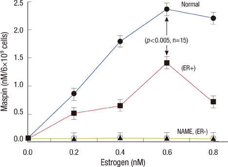
Figure 5 Eeffect of different amounts of estrogen on maspin synthesis in normal, estrogen receptor (ER)+, and ER- neutrophils. Neutrophil suspensions were prepared from the blood of patients with ER+ or ERbreast cancer receptor status and from age-matched normal female volunteers as described in the Methods section (n=15, in each group). The neutrophil preparations (6×10⁹ cells/L) were suspended in Hank's balanced salt solution (pH 7.4) and treated with different concentrations of estrogen (nM) as indicated. In a parallel set of experiments, neutrophil suspensions from age-matched normal female volunteers and from patients with ER+ or ER- breast cancer were incubated with NAME in the presence of varying amounts of estrogen. After incubation for 4 hours at 37℃, in vitro translation of maspin mRNAs was conducted and maspin synthesis in the reaction mixture was determined by enzyme linked immunosorbent assay as described in the Methods section. The solid circles (●) represent maspin synthesis in normal neutrophils, while the solid squares (■) indicate maspin synthesis in ER+ breast cancer neutrophils. The solid triangles (▲) represent maspin synthesis in ER- breast cancer neutrophils, while the hollow circles (◯) indicate maspin synthesis in the presence of NAME. Results are mean±SD of five different experiments in triplicate using blood of 15 patients with breast cancer and 15 normal female volunteers. NAME=L-NG Nitroarginine Methyl Ester.
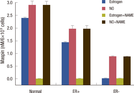
Figure 6 Effect of estrogen, nitric oxide (NO), and NAME on maspin synthesis in normal, estrogen receptor (ER)+, and ER- neutrophils. The neutrophil suspensions were prepared from the blood of patients with ER+ or ER- breast cancer and of age-matched normal female volunteers as described in the Methods section. The neutrophil preparations (6×10⁹ cells/L) were suspended in Hank's balanced salt solution (pH 7.4) and treated with estrogen (0.6 nM) and, NO (5 µM). In a separate experiment, a neutrophil suspension was incubated with NAME (10 mM) and with either NO or estrogen. Results are mean±SD of five different experiments in triplicate using blood of 15 patients with breast cancer and 15 normal female volunteers. NAME=L-NG Nitroarginine Methyl Ester.
Reference
-
1. Schneider HP, Jackisch C. Potential benefits of estrogens and progestogens on breast cancer. Int J Fertil Womens Med. 1998. 43:278–285.2. Beato M, Klug J. Steroid hormone receptors: an update. Hum Reprod Update. 2000. 6:225–236.
Article3. Zou Z, Anisowicz A, Hendrix MJ, Thor A, Neveu M, Sheng S, et al. Maspin, a serpin with tumor-suppressing activity in human mammary epithelial cells. Science. 1994. 263:526–529.
Article4. Hojo T, Akiyama Y, Nagasaki K, Maruyama K, Kikuchi K, Ikeda T, et al. Association of maspin expression with the malignancy grade and tumor vascularization in breast cancer tissues. Cancer Lett. 2001. 171:103–110.
Article5. Liu T, Pemberton PA, Robertson AD. Three-state unfolding and self-association of maspin, a tumor-suppressing serpin. J Biol Chem. 1999. 274:29628–29632.
Article6. Girish GV, Bhattacharya G, Sinha AK. The role of insulin dependent NO synthesis in the impaired production of maspin in human breast cancer. J Cancer Res Clin Oncol. 2006. 132:389–398.
Article7. Tlaskalová-Hogenová H, Stépánková R. Development of antibody formation in germ-free and conventionally reared rabbits: the role of intestinal lymphoid tissue in antibody formation to E. coli antigens. Folia Biol (Praha). 1980. 26:81–93.8. Cox RD, Frank CW. Determination of nitrate and nitrite in blood and urine by chemiluminescence. J Anal Toxicol. 1982. 6:148–152.
Article9. Klock JC, Bainton DF. Degranulation and abnormal bactericidal function of granulocytes procured by reversible adhesion to nylon wool. Blood. 1976. 48:149–161.
Article10. Cook L, Ross AM, Knight GB, Agnello V. Use of whole blood specimens for routine clinical quantitation of hepatitis C virus RNA does not increase assay sensitivity. J Clin Microbiol. 2000. 38:4326–4331.
Article11. Zimmerman R, Paluch U, Sprinzl M, Neupert W. Cell-free synthesis of the mitochondrial ADP/ATP carrier protein of Neurospora crassa. Eur J Biochem. 1979. 99:247–252.
Article12. Engvall E, Perlmann P. Enzyme-linked immunosorbent assay, Elisa. 3. Quantitation of specific antibodies by enzyme-labeled anti-immunoglobulin in antigen-coated tubes. J Immunol. 1972. 109:129–135.13. Motomura K, Ishitobi M, Komoike Y, Koyama H, Nagase H, Inaji H, et al. Expression of estrogen receptor beta and phosphorylation of estrogen receptor alpha serine 167 correlate with progression-free survival in patients with metastatic breast cancer treated with aromatase inhibitors. Oncology. 2010. 79:55–61.
Article14. Kahn NN, Sinha AK. Stimulation of prostaglandin E1 binding to human blood platelet membrane by insulin and the activation of adenylate cyclase. J Biol Chem. 1990. 265:4976–4981.
Article15. Scatchard G. The attractions of proteins for small molecules and ions. Ann N Y Acad Sci. 1949. 51:660–672.
Article16. Sakuma I, Stuehr DJ, Gross SS, Nathan C, Levi R. Identification of arginine as a precursor of endothelium-derived relaxing factor. Proc Natl Acad Sci U S A. 1988. 85:8664–8667.
Article17. Whiting KP, Restall CJ, Brain PF. Steroid hormone-induced effects on membrane fluidity and their potential roles in non-genomic mechanisms. Life Sci. 2000. 67:743–757.
Article18. Mauvais-Jarvis P, Kuttenn F, Gompel A. Antiestrogen action of progesterone in breast tissue. Breast Cancer Res Treat. 1986. 8:179–188.
Article19. Kiba T, Inamoto T, Nishimura T, Ueno M, Yanagihara K, Teramukai S, et al. The reversal of recurrence hazard rate between ER positive and negative breast cancer patients with axillary lymph node dissection (pathological stage I-III) 3 years after surgery. BMC Cancer. 2008. 8:323.
Article20. Rochefort H, Glondu M, Sahla ME, Platet N, Garcia M. How to target estrogen receptor-negative breast cancer? Endocr Relat Cancer. 2003. 10:261–266.
Article
- Full Text Links
-
- Actions
-
Cited
- CITED
-
- Close
- Share
-
- Similar articles
-
- Clinicopathological Significance of Maspin Expression in Breast Cancer
- Effect of Erythropoietin on the Production of Nitric Oxide in Trabecular Meshwork Cells
- Nitric Oxide Production in Mouse's Microglial Cells by Human Chorionic Gonadotropin
- Expression of Cyclooxygenase-2 in Human Breast Carcinoma: Relevance to Tumor Angiogenesis and Expression of Estrogen Receptor
- The Potential Role of Nitric Oxide in Halting Cancer Progression Through Chemoprevention
