The three-dimensional microstructure of trabecular bone: Analysis of site-specific variation in the human jaw bone
- Affiliations
-
- 1Department of Oral and Maxillofacial Radiology and Dental Research Institute, School of Dentistry, Seoul National University, Seoul, Korea. future3@snu.ac.kr
- 2Department of Oral and Maxillofacial Surgery, Ilsan Paik Hospital, Inje University College of Medicine, Goyang, Korea.
- 3A Plus Dental Clinic, Seoul, Korea.
- KMID: 2229604
- DOI: http://doi.org/10.5624/isd.2013.43.4.227
Abstract
- PURPOSE
This study was performed to analyze human maxillary and mandibular trabecular bone using the data acquired from micro-computed tomography (micro-CT), and to characterize the site-specific microstructures of trabeculae.
MATERIALS AND METHODS
Sixty-nine cylindrical bone specimens were prepared from the mandible and maxilla. They were divided into 5 groups by region: the anterior maxilla, posterior maxilla, anterior mandible, posterior mandible, and mandibular condyle. After the specimens were scanned using a micro-CT system, three-dimensional microstructural parameters such as the percent bone volume, bone specific surface, trabecular thickness, trabecular separation, trabecular number, structure model index, and degrees of anisotropy were analyzed.
RESULTS
Among the regions other than the condylar area, the anterior mandibular region showed the highest trabecular thickness and the lowest value for the bone specific surface. On the other hand, the posterior maxilla region showed the lowest trabecular thickness and the highest value for the bone specific surface. The degree of anisotropy was lowest at the anterior mandible. The condyle showed thinner trabeculae with a more anisotropic arrangement than the other mandibular regions.
CONCLUSION
There were microstructural differences between the regions of the maxilla and mandible. These results suggested that different mechanisms of external force might exist at each site.
Keyword
Figure
-
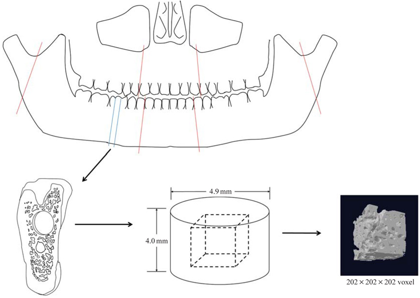
Fig. 1 Schematic description of specimen.
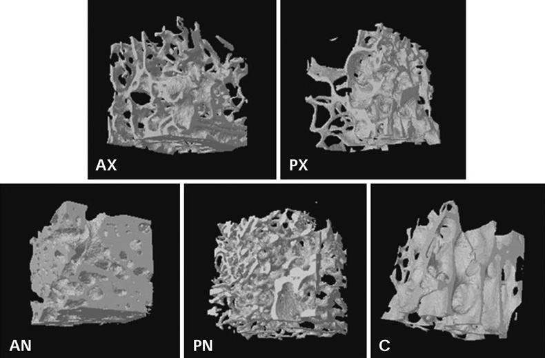
Fig. 2 Examples of 3D reconstruction of micro-CT data from bone specimens of specific regions.
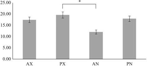
Fig. 3 Bone specific surface (BS/BV) of the various jaw regions.

Fig. 4 Trabecular thickeness (Tb.Th) of the various jaw regions.
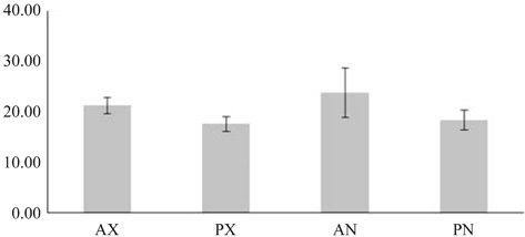
Fig. 5 Percent bone volume (BV/TV) of the various jaw regions.
Cited by 2 articles
-
Three-dimensional microstructure of human alveolar trabecular bone: a micro-computed tomography study
Ji-Hyun Lee, Hee-Jin Kim, Jeong-Ho Yun
J Periodontal Implant Sci. 2017;47(1):20-29. doi: 10.5051/jpis.2017.47.1.20.Comparison of alveolar ridge preservation methods using three-dimensional micro-computed tomographic analysis and two-dimensional histometric evaluation
Young-Seok Park, Sungtae Kim, Seung-Hee Oh, Hee-Jung Park, Sophia Lee, Tae-Il Kim, Young-Kyu Lee, Min-Suk Heo
Imaging Sci Dent. 2014;44(2):143-148. doi: 10.5624/isd.2014.44.2.143.
Reference
-
1. Lekholm U, Zarb GA. Patient selection and preparation. In : Brånemark PI, Zarb GA, Albrektsson T, editors. Tissue-integrated prostheses: osseointegration in clinical dentistry. Chicago: Quintessence;1985. p. 199–209.2. Ribeiro-Rotta RF, Lindh C, Rohlin M. Efficacy of clinical methods to assess jawbone tissue prior to and during endosseous dental implant placement: a systematic literature review. Int J Oral Maxillofac Implants. 2007; 22:289–300.3. Pothuaud L, Van Rietbergen B, Mosekilde L, Beuf O, Levitz P, Benhamou CL, et al. Combination of topological parameters and bone volume fraction better predicts the mechanical properties of trabecular bone. J Biomech. 2002; 35:1091–1099.
Article4. Thomsen JS, Ebbesen EN, Mosekilde L. Relationships between static histomorphometry and bone strength measurements in human iliac crest bone biopsies. Bone. 1998; 22:153–163.
Article5. Borah B, Dufresne TE, Cockman MD, Gross GJ, Sod EW, Myers WR, et al. Evaluation of changes in trabecular bone architecture and mechanical properties of minipig vertebrae by three-dimensional magnetic resonance microimaging and finite element modeling. J Bone Miner Res. 2000; 15:1786–1797.
Article6. Bassi F, Procchio M, Fava C, Schierano G, Preti G. Bone density in human dentate and edentulous mandibles using computed tomography. Clin Oral Implants Res. 1999; 10:356–361.
Article7. Norton MR, Gamble C. Bone classification: an objective scale of bone density using the computerized tomography scan. Clin Oral Implants Res. 2001; 12:79–84.
Article8. Weinstein RS, Hutson MS. Decreased trabecular width and increased trabecular spacing contribute to bone loss with aging. Bone. 1987; 8:137–142.
Article9. Hahn M, Vogel M, Pompesius-Kempa M, Delling G. Trabecular bone pattern factor - a new parameter for simple quantification of bone microarchitecture. Bone. 1992; 13:327–330.10. Fanuscu MI, Chang TL. Three-dimensional morphometric analysis of human cadaver bone: microstructural data from maxilla and mandible. Clin Oral Implants Res. 2004; 15:213–218.
Article11. Bryant SR. The effects of age, jaw site, and bone condition on oral implant outcomes. Int J Prosthodont. 1998; 11:470–490.12. Misch CE, Qu Z, Bidez MW. Mechanical properties of trabecular bone in the human mandible: implications for dental implant treatment planning and surgical placement. J Oral Maxillofac Surg. 1999; 57:700–708.
Article13. Ulm CW, Kneissel M, Hahn M, Solar P, Matejka M, Donath K. Characteristics of the cancellous bone of edentulous mandibles. Clin Oral Implants Res. 1997; 8:125–130.
Article14. Yi WJ, Heo MS, Lee SS, Choi SC, Huh KH. Comparison of trabecular bone anisotropies based on fractal dimensions and mean intercept length determined by principal axes of inertia. Med Biol Eng Comput. 2007; 45:357–364.
Article15. Ulrich D, van Rietbergen B, Laib A, Rüegsegger P. The ability of three-dimensional structural indices to reflect mechanical aspects of trabecular bone. Bone. 1999; 25:55–60.
Article16. Jensen O. Site classification for the osseointegrated implant. J Prosthet Dent. 1989; 61:228–234.
Article17. Friberg B, Sennerby L, Roos J, Lekholm U. Identification of bone quality in conjunction with insertion of titanium implants. A pilot study in jaw autopsy specimens. Clin Oral Implants Res. 1995; 6:213–219.
Article18. Riggs BL, Hodgson SF, O'Fallon WM, Chao EY, Wahner HW, Muhs JM, et al. Effect of fluoride treatment on the fracture rate in postmenopausal women with osteoporosis. N Engl J Med. 1990; 322:802–809.
Article19. Koh KJ, Park HN, Kim KA. Prediction of age-related osteoporosis using fractal analysis on panoramic radiographs. Imaging Sci Dent. 2012; 42:231–235.
Article20. Amer ME, Heo MS, Brooks SL, Benavides E. Anatomical variations of trabecular bone structure in intraoral radiographs using fractal and particles count analyses. Imaging Sci Dent. 2012; 42:5–12.
Article21. Giesen EB, van Eijden TM. The three-dimensional cancellous bone architecture of the human mandibular condyle. J Dent Res. 2000; 79:957–963.
Article22. Giesen EB, Ding M, Dalstra M, van Eijden TM. Mechanical properties of cancellous bone in the human mandibular condyle are anisotropic. J Biomech. 2001; 34:799–803.
Article23. van Eijden TM, van der Helm PN, van Ruijven LJ, Mulder L. Structural and mechanical properties of mandibular condylar bone. J Dent Res. 2006; 85:33–37.
Article24. van Ruijven LJ, Giesen EB, van Eijden TM. Mechanical significance of the trabecular microstructure of the human mandibular condyle. J Dent Res. 2002; 81:706–710.
Article25. Moon HS, Won YY, Kim KD, Ruprecht A, Kim HJ, Kook HK, et al. The three-dimensional microstructure of the trabecular bone in the mandible. Surg Radiol Anat. 2004; 26:466–473.
Article
- Full Text Links
-
- Actions
-
Cited
- CITED
-
- Close
- Share
-
- Similar articles
-
- Changes in Microstructure of Proximal Femoral Trabecular Bone
- Three-dimensional microstructure of human alveolar trabecular bone: a micro-computed tomography study
- Measurement of Trabecular Bone Parameters in Porcine Vertebral Bodies Using Multidetector CT: Evaluation of Reproducibility of 3-Dimensional CT Histomorphometry
- Femoral bone structure in Otsuka Long-Evans Tokushima Fatty rats
- Differences in the effects of BMI on bone microstructure between loaded and unloaded bones assessed by HR-pQCT in Japanese postmenopausal women
