Korean Circ J.
2009 Sep;39(9):352-358. 10.4070/kcj.2009.39.9.352.
Cardiac Functional Evaluation Using Vector Velocity Imaging After Chemotherapy Including Anthracyclines in Children With Cancer
- Affiliations
-
- 1Department of Pediatrics, Keimyung University School of Medicine, Daegu, Korea. kimyhped@hanmail.net
- 2Department of Pediatrics, School of Medicine, Kyungpook National University, Daegu, Korea.
- KMID: 2225670
- DOI: http://doi.org/10.4070/kcj.2009.39.9.352
Abstract
- BACKGROUND AND OBJECTIVES
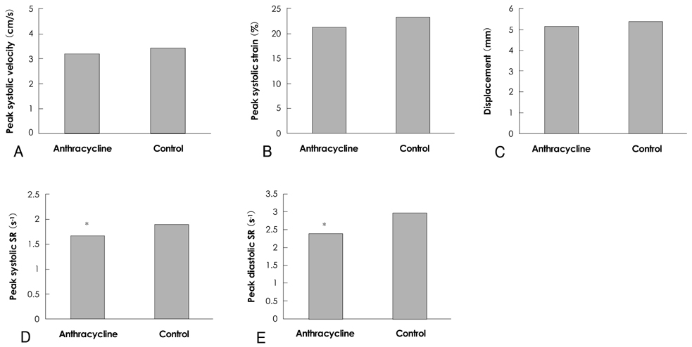
Anthracyclines are effective drugs that are widely used in pediatric cancer treatment. Previous studies have demonstrated that exposure to low-dose anthracyclines (<300 mg/m2) induces a progressive decrease in cardiac function during long-term follow-up. The goal of this study was to assess left ventricular function using vector velocity imaging (VVI) in children undergoing low-dose anthracycline therapy. SUBJECTS AND METHODS: We examined 14 asymptomatic patients who had been treated with anthracyclines and had normal fractional shortening (FS) and ejection fraction (EF). In all of the patients, standard two-dimensional (2D) pulsed and tissue Doppler echocardiographic measurements were taken from an apical 4-chamber view. The peak myocardial velocity, peak strain rate (SR), peak strain, and displacement were obtained from VVI. Data were compared with 14 age-matched healthy controls. RESULTS: From the regional wall motion analysis using VVI in the left ventricle, the peak myocardial velocity and displacement of the lateral wall were increased significantly more than the septum, and there were no significant differences between the patients and the controls. Although systolic strain, and the systolic and diastolic SRs showed no significant differences between the septum and lateral wall in the controls, those of septum, in the patients, were decreased significantly more than those of lateral wall (p<0.05). In comparison with the controls, these changes in septal strain and SRs of patients were significant (p<0.05). CONCLUSION: Anthracycline therapy, even low-dose, can induce changes in regional wall function before global dysfunction. Also, the strain and SR obtained from VVI may be useful for early detection of these changes.
Keyword
MeSH Terms
Figure
Reference
-
1. Singal PK, Iliskovic N. Doxorubicin-induced cardiomyopathy. N Engl J Med. 1998. 339:900–905.2. Nysom K, Holm K, Lipsitz SR, et al. Relationship between cumulative anthracycline dose and late cardiotoxicity in childhood acute lymphoblastic leukemia. J Clin Oncol. 1998. 16:545–550.3. Lipshultz SE, Lipsitz SR, Sallan SE, et al. Chronic progressive cardiac dysfunction years after doxorubicin therapy for childhood acute lymphoblastic leukemia. J Clin Oncol. 2005. 23:2629–2636.4. Ganame J, Claus P, Uyttebroeck A, et al. Myocardial dysfunction late after low-dose anthracycline treatment in asymptomatic pediatric patients. J Am Soc Echocardiogr. 2007. 20:1351–1358.5. Haq MM, Legha SS, Choksi J, et al. Doxorubicin-induced congestive heart failure in adults. Cancer. 1985. 56:1361–1365.6. Steinherz LJ, Graham T, Hurwitz R, et al. Guidelines for cardiac monitoring of children during and after anthracycline therapy: report of the Cardiology Committee of the Childrens Cancer Study Group. Pediatrics. 1992. 89:942–949.7. Pirat B, McCulloch ML, Zoghbi WA. Evaluation of global and regional right ventricular systolic function in patients with pulmonary hypertension using a novel speckle tracking method. Am J Cardiol. 2006. 98:699–704.8. Pislaru C, Abraham TP, Belohlavek M. Strain and strain rate echocardiography. Curr Opin Cardiol. 2002. 17:443–454.9. Lipshultz SE, Colan SD, Gelber RD, Perez-Atayde AR, Sallan SE, Sanders SP. Late cardiac effects of doxorubicin therapy for acute lymphoblastic leukemia in childhood. N Engl J Med. 1991. 324:808–815.10. Lipshultz SE, Lipsitz SR, Mone SM, et al. Female sex and drug dose as risk factors for late cardiotoxic effects of doxorubicin therapy for childhood cancer. N Engl J Med. 1995. 332:1738–1743.11. Nysom K, Colan SD, Lipshultz SE. Late cardiotoxicity following anthracycline therapy for childhood cancer. Prog Pediatr Cardiol. 1998. 8:121–128.12. Yeung ST, Yoong C, Spink J, Galbraith A, Smith PJ. Functional myocardial impairment in children treated with anthracyclines for cancer. Lancet. 1991. 337:816–818.13. Dorup I, Levitt G, Sullivan I, Sorensen K. Prospective longitudinal assessment of late anthracycline cardiotoxicity after childhood cancer: the role of diastolic function. Heart. 2004. 90:1214–1216.14. Stoddard MF, Seeger J, Liddell NE, Hadley TJ, Sullivan DM, Kupersmith J. Prolongation of isovolumetric relaxation time as assessed by Doppler echocardiography predicts doxorubicin-induced systolic dysfunction in humans. J Am Coll Cardiol. 1992. 20:62–69.15. Eidem BW, Sapp BG, Suarez CR, Cetta F. Usefulness of the myocardial performance index for early detection of anthracycline-induced cardiotoxicity in children. Am J Cardiol. 2001. 87:1120–1122.16. Kapusta L, Thijssen JM, Groot-Loonen J, Antonius T, Mulder J, Daniels O. Tissue Doppler imaging in detection of myocardial dysfunction in survivors of childhood cancer treated with anthracyclines. Ultrasound Med Biol. 2000. 26:1099–1108.17. Cho KI, Lee SH, Jang SH, Lee DW, Lee HG, Kim TI. Assessment of left atrial function and remodeling in patients with atrial fibrillation by performing strain echocardiography: a prospective study to assess the influence of renin-angiotensin system inhibitors on atrial fibrillation. Korean Circ J. 2008. 38:305–312.18. Choi JO, Cho SJ, Yang JH, et al. Systolic long axis function of the left ventricle, as assessed by 2-D strain, is reduced in the patients who have diastolic dysfunction and a normal ejection fraction. Korean Circ J. 2008. 38:250–256.19. Cottin Y, Touzery C, Coudert B, et al. Diastolic or systolic left and right ventricular impairment at moderate doses of anthracycline?: a 1-year follow-up study of women. Eur J Nucl Med. 1996. 23:511–516.
- Full Text Links
- Actions
-
Cited
- CITED
-
- Close
- Share
- Similar articles
-
- Long-term cardiac composite risk following adjuvant treatment in breast cancer patients
- Pathophysiology and preventive strategies of anthracycline-induced cardiotoxicity
- Follow-up Study of Children with Anthracycline Cardiotoxicity
- Vascular endothelial dysfunction after anthracyclines treatment in children with acute lymphoblastic leukemia
- MRI for the Functional Evaluation of Systemic Right Ventricle in TGA


