Korean Circ J.
2010 Jun;40(6):260-265. 10.4070/kcj.2010.40.6.260.
The Impact of Mitral Annular Calcification on Left Ventricular Function in Nonagenarians
- Affiliations
-
- 1Division of Cardiology, Heart Research Institute, College of Medicine, Chung-Ang University, Seoul, Korea. swivus@gmail.com
- KMID: 2225185
- DOI: http://doi.org/10.4070/kcj.2010.40.6.260
Abstract
- BACKGROUND AND OBJECTIVES
Mitral annular calcification (MAC) is known to be associated with degenerative processes of the cardiac fibrous skeleton and cardiovascular disease mortality. However, MAC has not been evaluated in an extreme age group (patients > or =90 years of age). In this study, the clinical significance of MAC associated with aging was examined in this age group and compared with MAC associated with aging in a younger (20 to 50 years of age) group of patients.
SUBJECTS AND METHODS
We assessed echocardiographic parameters in 43 nonagenarians and 51 young patients. In the nonagenarian group, patient's age was 92+/-2 years and 27% were male; in the young control group, patient's age was 36+/-9 years and 51% were male. Comprehensive M-mode and Doppler echocardiography, including tissue Doppler imaging, were performed. The frequency and severity of MAC was assessed from the leading anterior to the trailing posterior edge at its largest width for least 3 cardiac cycles.
RESULTS
Echocardiography showed that the left ventricular (LV) end-diastolic dimension was larger in the young controls (p=0.007); however, the ejection fraction (EF) was lower in the nonagenarian group (p=0.001). The frequency of MAC was greater in nonagenarians {42/43 (97%)} than in controls {9/51 (17%), p<0.0001}. The maximal width of MAC was larger in nonagenarians (0.52+/-0.17 mm and 0.05+/-0.13 mm, p<0.0001). MAC was correlated with LV mass index (g/m2) (r=0.280, p=0.014) and EF (%) (r=-0.340, p=0.001). More importantly, early mitral inflow velocity/early diastolic mitral annulus velocity (E/E') was strongly correlated with MAC in non-agenarians (r= 0.683, p<0.0001).
CONCLUSION
MAC may be associated with extreme age and increased LV filling pressure in nonagenarians. Further study is necessary to assess the cardiovascular mortality and structural changes related to mitral annulus calcification associated with aging.
Keyword
MeSH Terms
Figure
-

Fig. 1 The measurement of mitral annulus calcification (MAC) in a nonagenarian. The thickest measured width of MAC was 7.6 mm at the apical four chamber view.

Fig. 2 The frequency and severity of mitral annulus calcification (MAC) were higher in nonagenarians. A: frequency of MAC. B: MAC width.
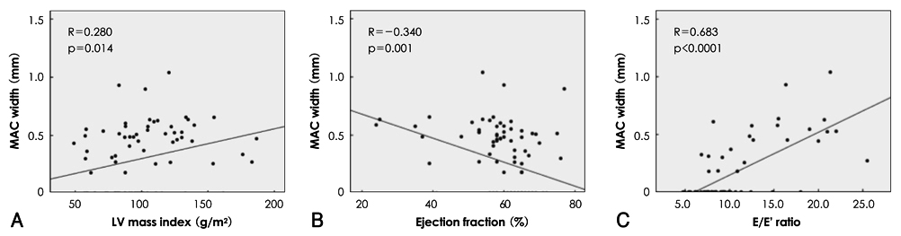
Fig. 3 The correlations of mitral annular calcification (MAC) width. A: left ventricular mass index. B: ejection fraction. C: E/E' ratio.
Reference
-
1. D'Cruz I, Panetta F, Cohen H, Glick G. Submitral calcification or sclerosis in elderly patients: M-mode and two-dimensional echocardiography in "mitral annulus calcification". Am J Cardiol. 1979. 44:31–38.2. Hirschfeld DS, Emilson BB. Echocardiogram in calcified mitral annulus. Am J Cardiol. 1975. 36:354–356.3. Roberts WC. The senile cardiac calcification syndrome. Am J Cardiol. 1986. 58:572–574.4. Pomerance A. Pathology of the heart with and without cardiac failure in the aged. Br Heart J. 1965. 27:697–710.5. Burnside JW, Desanctis RW. Bacterial endocarditis on calcification of the mitral annulus fibrosus. Ann Intern Med. 1972. 76:615–618.6. Jeon DS, Atar S, Brasch AV, et al. Association of mitral annulus calcification, aortic valve sclerosis and aortic root calcification with abnormal myocardial perfusion single photon emission tomography in subjects age < or =65 years old. J Am Coll Cardiol. 2001. 38:1988–1993.7. Tenenbaum A, Shemesh J, Fisman EZ, Motro M. Advanced mitral annular calcification is associated with severe coronary calcification on fast dual spiral computed tomography. Invest Radiol. 2000. 35:193–198.8. Adler Y, Herz I, Vaturi M, et al. Mitral annular calcium detected by transthoracic echocardiography is a marker for high prevalence and severity of coronary artery disease in patients undergoing coronary angiography. Am J Cardiol. 1998. 82:1183–1186.9. Aronow WS, Ahn C, Kronzon I. Association of mitral annular calcium and of aortic cuspal calcium with coronary artery disease in older patients. Am J Cardiol. 1999. 84:1084–1085.10. Gardin JM, McClelland R, Kitzman D, et al. M-mode echocardiographic predictors of six- to seven-year incidence of coronary heart disease, stroke, congestive heart failure, and mortality in an elderly cohort (the Cardiovascular Health Study). Am J Cardiol. 2001. 87:1051–1057.11. D'Cruz IA, Cohen HC, Prabhu R, Bisla V, Glick G. Clinical manifestations of mitral annulus calcification, with emphasis on its echocardiographic features. Am Heart J. 1977. 94:367–377.12. Mellino M, Salcedo EE, Lever HM, Vasudevan G, Kramer JR. Echographic-quantified severity of mitral annulus calcification: prognostic correlation to related hemodynamic, valvular, rhythm, and conduction abnormalities. Am Heart J. 1982. 103:222–225.13. Fox CS, Vasan RS, Parise H, et al. Mitral annular calcification predicts cardiovascular morbidity and mortality: the Framingham Heart Study. Circulation. 2003. 107:1492–1496.14. Adler Y, Koren A, Fink N, et al. Association between mitral annulus calcification and carotid atherosclerotic disease. Stroke. 1998. 29:1833–1837.15. Yang SK, Jo KR. 2008 Statistic on the aged in Korea. 2008. Korea National Statistical Office.16. Lang RM, Bierig M, Devereux RB, et al. Recommendations for chamber quantification: a report from the American Society of Echocardiography's Guidelines and Standards Committee and the Chamber Quantification Writing Group, developed in conjunction with the European Association of Echocardiography, a branch of the European Society of Cardiology. J Am Soc Echocardiogr. 2005. 18:1440–1463.17. Devereaux RB, Alonso DR, Lutas EM, et al. Echocardiographic assessment of left ventricular hypertrophy: comparison to necropsy findings. Am J Cardiol. 1986. 57:450–458.18. Sadig A, Choudhury M, Ali K, et al. Echocardiographic characteristics in patients >100 years of age. Am J Cardiol. 2007. 100:1792–1794.19. Wong RC, Yip JW, Gupta A, Yang H, Ling LH. Echocardiographic left ventricular mass in a multiethnic Southeast Asian population: proposed new gender and age-specific norms. Echocardiography. 2008. 25:805–811.20. Park SH, Chung NS, Cho SY, Shin DH, Kim SI. Quantative analysis of mitral valvular calcification in mitral stenosis. Korean Circ J. 1994. 24:38–52.21. Park KS, Yoh KG, Cho YK, Yoon JH, Choi KH. The effects of excimer laser coronary angioplasty in calcified lesions: investigation with intravascular ultrasound. Korean Circ J. 1994. 24:609–616.22. Lee YG, Park KS, Song KS, et al. Coronary artery calcification its incidence and significance in patients detected by cineangiography. Korean Circ J. 1994. 24:646–652.23. Mintz GS, Popma JJ, Pichard AD, et al. Patterns of calcification in coronary artery disease: a statistical analysis of intravascular ultrasound and coronary angiography in 1155 lesions. Circulation. 1995. 91:1959–1965.24. Rhew JY, Jeong MH, Kang KT, et al. A giant aneurysm of the sinus of Valsalva with calcification. Korean Circ J. 2001. 31:114–118.25. Kim SH, Woo SY, Oh YK, et al. A case of recurrent thrombus associated with left atrial calcification. Korean Circ J. 2004. 34:323–327.26. Han SH, Park CG, Park SW, et al. High aortic stiffness assessed by pulse wave velocity is an independent predictor of coronary artery calcification and stenosis in suspected coronary artery disease patients. Korean Circ J. 2004. 34:468–476.27. Boon A, Cheriex E, Lodder J, Kessels F. Cardiac valve calcification: characteristics of patients with calcification of the mitral annulus or aortic valve. Heart. 1997. 78:472–474.28. Rao AK, Djamali A, Korcarz CE, Aeschlimann SE, Wolff MR, Stein JH. Mitral annular calcification is associated with reduced left ventricular function and inflammation in patients with chronic kidney disease. J Am Soc Echocardiogr. 2008. 21:747–750.29. Lim YH, Lee JU, Kim KS, et al. Association between inappropriateness of left ventricular mass and left ventricular diastolic dysfunction: a study using the tissue doppler parameter, E/E'. Korean Circ J. 2009. 39:138–144.30. Nagueh SF, Kopelen HA, Quinones M. Assessment of left ventricular filling pressures by Doppler in the presence of atrial fibrillation. Circulation. 1996. 94:2138–2145.
- Full Text Links
-
- Actions
-
Cited
- CITED
-
- Close
- Share
-
- Similar articles
-
- Mitral Valve Replacement for Bulky, Calcified Mitral Annulus: A Case Report
- Modified Surgical Intervention for Extensive Mitral Valve Endocarditis and Posterior Mitral Annular Calcification
- Assessment of Diastolic Function Using Mitral Annulus Velocity by Doppler Tissue Velocity in the Patients with Left Ventricular Hypertrophy
- Two Cases of Caseous Calcification of the Mitral Annulus
- Mitral Valve Repair for Barlow’s Disease with Mitral Annular and Subvalvular Calcification: A Case Report
