A Case of a Pulmonary Arteriovenous Malformation With Ebstein's Anomaly
- Affiliations
-
- 1Department of Internal Medicine, Kangdong Sacred Heart Hospital, Seoul, Korea.
- 2Department of Internal Medicine and Sejong Medical Research Institute, Sejong General Hospital, Bucheon, Korea. masque@sejongh.co.kr
- KMID: 2225170
- DOI: http://doi.org/10.4070/kcj.2010.40.12.684
Abstract
- A pulmonary arteriovenous malformation (PAVM) is a rare pulmonary vascular anomaly presenting as dyspnea or recurrent epistaxis. Ebstein's anomaly (EA), a congenital cardiac malformation, is also a rare condition. There have been no reports concerning the co-existence of PAVM with hereditary hemorrhagic telangiectasia (HHT) and EA. A 40-year-old woman was admitted with a 2-month history of increasing dyspnea and several years of recurrent epistaxis. On transthoracic echocardiography, she was diagnosed with EA and agreed to undergo surgical treatment. A chest CT angiography showed a 12-mm serpiginous vascular structure suspicious for a PAVM and a liver CT suggested HTT. Although it is unclear whether or not a concurrent PAVM and EA have an embryologic or genetic relationship, we report a case of a PAVM with EA. Further genetic and embryonic studies are needed to identify a possible relationship of the two medical conditions.
Keyword
MeSH Terms
Figure
-
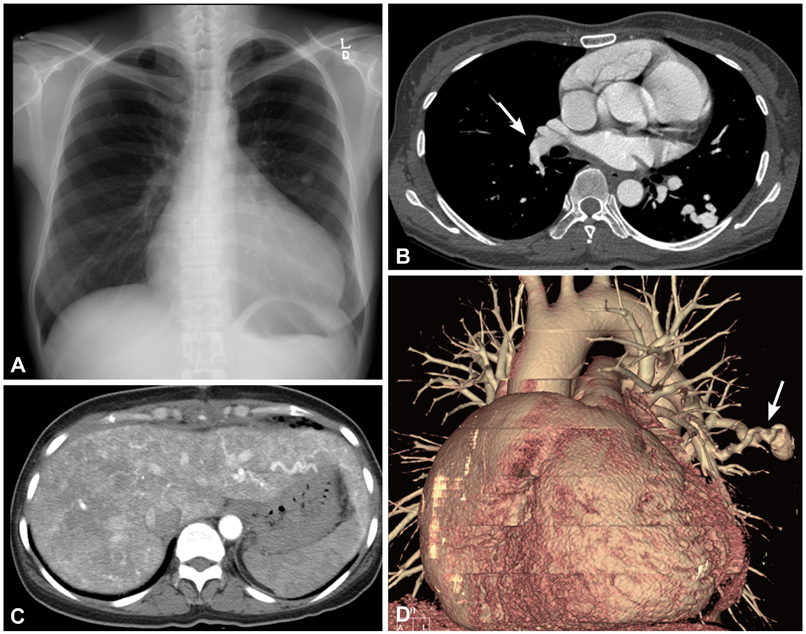
Fig. 1 A: initial chest X-ray showing a round nodular opacity on the left lung. B and D: on the chest CT angiography, a pulmonary arteriovenous malformation was detected in the left lower lobe (arrows). C: the arterial phase of the liver CT revealed a severe tortuous dilatation of hepatic arteries and early visualization of hepatic veins with multifocal arteriovenous malformations.
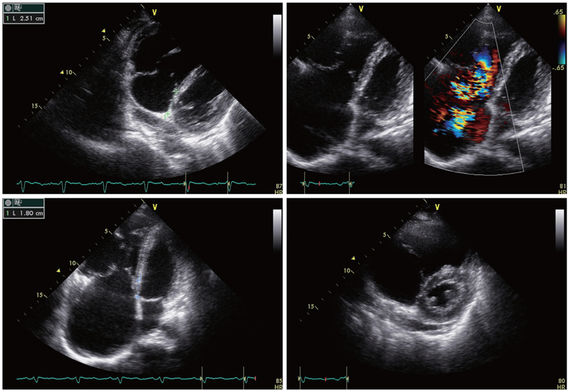
Fig. 2 Transthoracic echocardiography: Ebstein's anomaly. The displacement index was mesured (15.6 mm/m2). Tethering of the septal and posterior leaflets of the tricuspid valve was observed with central coaptation failure and severe regurgitation. A portion of the right ventricle was atrialized because of apical displacement of the tricuspid valve. The right atrium and ventricle were markedly enlarged.
Reference
-
1. Pick A, Deschamps C, Stanson AW. Pulmonary arteriovenous fistula: presentation, diagnosis, and treatment. World J Surg. 1999. 23:1118–1122.2. Khurshid I, Downie GH. Pulmonary arteriovenous malformation. Postgrad Med J. 2002. 78:191–197.3. Jang HJ, Kim MS, Kim SY, et al. A case of embolization seen in pulmonary arteriovenous malformation in a patient with Osler-Rendu-Weber syndrome. Korean Circ J. 2006. 36:820–822.4. Gossage JR, Kanj G. Pulmonary arteriovenous malformations: a state of the art review. Am J Respir Crit Care Med. 1998. 158:643–661.5. Swanson KL, Prakash UB, Stanson AW. Pulmonary arteriovenous fistulas: Mayo clinic experience, 1982-1997. Mayo Clin Proc. 1999. 74:671–680.6. Memeo M, Scardapane A, De Blasi R, Sabba C, Carella A, Angelelli G. Diagnostic imaging in the study of visceral involvement of hereditary haemorrhagic telangiectasia. Radiol Med. 2008. 113:547–566.7. Attenhofer Jost CH, Connolly HM, Dearani JA, Edwards WD, Danielson GK. Ebstein's anomaly. Circulation. 2007. 115:277–285.8. Attenhofer Jost CH, Connolly HM, Edwards WD, Hayes D, Warnes CA, Danielson GK. Ebstein's anomaly: review of a multifaceted congenital cardiac condition. Swiss Med Wkly. 2005. 135:269–281.9. Andelfinger G, Wright KN, Lee HS, Siemens LM, Benson DW. Canine tricuspid valve malformation, a model of human Ebstein anomaly, maps to dog chromosome 9. J Med Genet. 2003. 40:320–324.10. Marchuk DA. The molecular genetics of hereditary hemorrhagic telangiectasia. Chest. 1997. 111:6 Suppl. 79S–82S.11. Kim MA, Cho SW, Lee WS, et al. Ebastein's anomaly in adults. Korean Circ J. 1988. 18:673–680.12. Shovlin CL, Guttmacher AE, Buscarini E, et al. Diagnostic criteria for hereditary hemorrhagic telangiectasia (Rendu-Osler-Weber syndrome). Am J Med Genet. 2000. 91:66–67.
- Full Text Links
-
- Actions
-
Cited
- CITED
-
- Close
- Share
-
- Similar articles
-
- A Case of Multiple Pulmonary Arteriovenous Malformation Treated with Coil Embolization
- Cone Repair in Adult Patients with Ebstein Anomaly
- Ebstein's Anomaly in a 74-year-old Man
- A Case of Cerebellar Infarction with Pulmonary Arteriovenous Malformation
- A Case of the Bronchial Artery-Pulmonary Vein Malformation
