Korean Circ J.
2011 Mar;41(3):137-142. 10.4070/kcj.2011.41.3.137.
Determination of Diastolic Dysfunction Cut-Off Value by Tissue Doppler Imaging in Adults 70 Years of Age or Older: A Comparative Analysis of Pulsed-Wave and Color-Coded Tissue Doppler Imaging
- Affiliations
-
- 1Department of Cardiology, Dong-A University College of Medicine, Busan, Korea. thpark65@dau.ac.kr
- KMID: 2225127
- DOI: http://doi.org/10.4070/kcj.2011.41.3.137
Abstract
- BACKGROUND AND OBJECTIVES
The cut-off value of diastolic dysfunction by tissue Doppler imaging (TDI) is affected by aging and modalities used (pulsed-wave vs. color-coded). The purpose of this study was to investigate the diastolic function of healthy elderly people and to determine the appropriate cut-off value of diastolic dysfunction in elderly individuals.
SUBJECTS AND METHODS
Healthy volunteers (n=76) and patients with hypertension (n=51) aged > or =70 years underwent 2-dimensional and Doppler echocardiography. Mitral annulus velocities of TDI were measured at septal and lateral sites using the pulsed-wave and color-coded modalities. The appropriate cut-off value of diastolic dysfunction for healthy elderly individuals was defined as the lower limit of the 95% confidence interval for early diastolic mitral annulus velocity (Ea).
RESULTS
The mean septal and lateral Ea were 6.5+/-1.5 and 8.3+/-1.7 cm/s, respectively, by pulsed-wave TDI, and 6.1+/-1.4 and 7.9+/-1.7 cm/s, respectively, by color-coded TDI. The cut-off values for diastolic dysfunction were as follows: septal and lateral Ea were 6.1 and 7.9 cm/s by pulsed-wave TDI, and 5.7 and 7.5 cm/s by color-coded TDI, respectively. When the group was stratified by gender, Ea was significantly lower in women than men.
CONCLUSION
When interpreting diastolic function as measured by TDI in elderly subjects, different cut-off values should be considered based on the TDI modality, annulus site, and gender.
Keyword
MeSH Terms
Figure
-
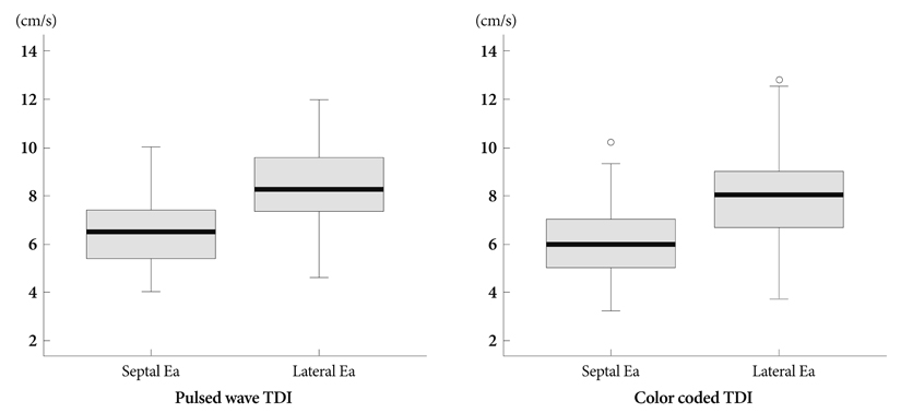
Fig. 1 Box plots showing distributions of septal and lateral Ea measured by pulsed-wave TDI and color-coded TDI, respectively. Box heights reflect interquartile ranges (IQRs). Vertical capped lines (whiskers) extend to most extreme data points within 1.5 IQRs of boxes. Data beyond 1.5 IQRs from boxes are plotted individually. Ea: early diastolic mitral annulus velocity, TDI: tissue Doppler imaging.
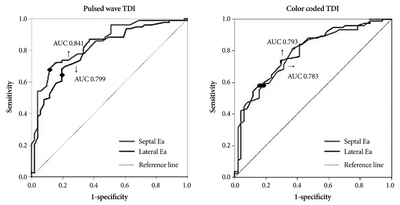
Fig. 2 Receiver-operator characteristic curve of Ea. The point (●) is the sensitivity and specificity of cut-off value, which is defined by the lower limit of the 95% CI for Ea to detect hypertensive patients who have diastolic dysfunction. TDI: tissue Doppler imaging.
Reference
-
1. Nishimura RA, Tajik AJ. Evaluation of diastolic filling of left ventricle in health and disease: Doppler echocardiography is the clinician's Rosetta Stone. J Am Coll Cardiol. 1997. 30:8–18.2. Yamada H, Oki T, Mishiro Y, et al. Effect of aging on diastolic left ventricular myocardial velocities measured by pulsed tissue Doppler imaging in healthy subjects. J Am Soc Echocardiogr. 1999. 12:574–581.3. Rodriguez L, Garcia M, Ares M, Griffin BP, Nakatani S, Thomas JD. Assessment of mitral annular dynamics during diastole by Doppler tissue imaging: comparison with mitral Doppler inflow in subjects without heart disease and in patients with left ventricular hypertrophy. Am Heart J. 1996. 131:982–987.4. Hirota Y. A clinical study of left ventricular relaxation. Circulation. 1980. 62:756–763.5. Garcia MJ, Rodriguez L, Ares M, Griffin BP, Thomas JD, Klein AL. Differentiation of constrictive pericarditis from restrictive cardiomyopathy: assessment of left ventricular diastolic velocities in longitudinal axis by Doppler tissue imaging. J Am Coll Cardiol. 1996. 27:108–114.6. Zarich SW, Kowalchuk GJ, McGuire MP, Benotti PN, Mascioli EA, Nesto RW. Left ventricular filling abnormalities in asymptomatic morbid obesity. Am J Cardiol. 1991. 68:377–381.7. Wong CY, O'Moore-Sullivan T, Leano R, Byrne N, Beller E, Marwick TH. Alterations of left ventricular myocardial characteristics associated with obesity. Circulation. 2004. 110:3081–3087.8. Moon CI, Choi JW, Cho YB, Shin WY, Song CS. Assessment of normal mitral annulus velocity using Doppler tissue imaging. Korean Circ J. 2001. 31:662–669.9. Schiller NB, Shah PM, Crawford M, et al. Recommendations for quantitation of the left ventricle by two-dimensional echocardiography. American Society of Echocardiography Committee on Standards, Subcommittee on Quantitation of Two-Dimensional Echocardiograms. J Am Soc Echocardiogr. 1989. 2:358–367.10. Lang RM, Bierig M, Devereux RB, et al. Recommendations for chamber quantification. Eur J Echocardiogr. 2006. 7:79–108.11. Hunt SA. ACC/AHA 2005 guideline update for the diagnosis and management of chronic heart failure in the adult: a report of the American College of Cardiology/American Heart Association Task Force on Practice Guidelines (Writing Committee to Update the 2001 Guidelines for the Evaluation and Management of Heart Failure). J Am Coll Cardiol. 2005. 46:e1–e82.12. Ren X, Ristow B, Na B, Ali S, Schiller NB, Whooley MA. Prevalence and prognosis of asymptomatic left ventricular diastolic dysfunction in ambulatory patients with coronary heart disease. Am J Cardiol. 2007. 99:1643–1647.13. Ha JW, Oh JK. The pathophysiology and diagnostic approaches for diastolic left ventricular dysfunction: a clinical perspective. Korean Circ J. 2005. 35:865–876.14. Redfield MM, Jacobsen SJ, Burnett JC Jr, Mahoney DW, Bailey KR, Rodeheffer RJ. Burden of systolic and diastolic ventricular dysfunction in the community: appreciating the scope of the heart failure epidemic. JAMA. 2003. 289:194–202.15. Nagueh SF. Search for non-invasive load-independent indices of left ventricular relaxation. Clin Sci. 2003. 105:395–397.16. Sohn DW, Chai IH, Lee DJ, et al. Assessment of mitral annulus velocity by Doppler tissue imaging in the evaluation of left ventricular diastolic function. J Am Coll Cardiol. 1997. 30:474–480.17. Garcia MJ, Thomas JD, Klein AL. New Doppler echocardiographic applications for the study of diastolic function. J Am Coll Cardiol. 1998. 32:865–875.18. Nagueh SF, Middleton KJ, Kopelen HA, Zoghbi WA, Quiñones MA. Doppler tissue imaging: a noninvasive technique for evaluation of left ventricular relaxation and estimation of filling pressures. J Am Coll Cardiol. 1997. 30:1527–1533.19. Chetboul V, Escriou C, Tessier D, et al. Tissue Doppler imaging detects early asymptomatic myocardial abnormalities in a dog model of Duchenne's cardiomyopathy. Eur Heart J. 2004. 25:1934–1939.20. Strand A, Kjeldsen SE, Gudmundsdottir H, Os I, Smith G, Bjørnerheim R. Tissue Doppler imaging describes diastolic function in men prone to develop hypertension over twenty years. Eur J Echocardiogr. 2008. 9:34–39.21. Munagala VK, Jacobsen SJ, Mahoney DW, Rodeheffer RJ, Bailey KR, Redfield MM. Association of newer diastolic function parameters with age in healthy subjects: a population-based study. J Am Soc Echocardiogr. 2003. 16:1049–1056.22. Anderson B. Echocardiography: the normal examination and echocardiographic measurements. 2007. 2nd ed. Australia: MGA graphics;323–327.23. Masoudi FA, Havranek EP, Smith G, et al. Gender, age, and heart failure with preserved left ventricular systolic function. J Am Coll Cardiol. 2003. 41:217–223.24. Galvao M, Kalman J, DeMarco T, et al. Gender differences in in-hospital management and outcomes in patients with decompensated heart failure: analysis from the Acute Decompensated Heart Failure National Registry (ADHERE). J Card Fail. 2006. 12:100–107.25. Yancy CW, Lopatin M, Stevenson LW, De Marco T, Fonarow GC. Clinical presentation, management, and in-hospital outcomes of patients admitted with acute decompensated heart failure with preserved systolic function: a report from the Acute Decompensated Heart Failure National Registry (ADHERE) Database. J Am Coll Cardiol. 2006. 47:76–84.26. Redfield MM, Jacobsen SJ, Borlaug BA, Rodeheffer RJ, Kass DA. Age- and gender-related ventricular-vascular stiffening: a community-based study. Circulation. 2005. 112:2254–2262.
- Full Text Links
-
- Actions
-
Cited
- CITED
-
- Close
- Share
-
- Similar articles
-
- Myocardial Function in Infant Kawasaki Disease with Tissue Doppler Imaging
- Doppler ultrasonography of the lower extremity arteries: anatomy and scanning guidelines
- Assessment of Normal Mitral Annulus Velocity by Doppler Tissue Imaging
- Tissue Doppler Imaging for the Assessment of Left Ventricular Diastolic Function
- Visualization of Coronary Arteries by Color-Coded Transesophageal Doppler Echocardiography
