Korean Circ J.
2015 Nov;45(6):500-509. 10.4070/kcj.2015.45.6.500.
A Novel Echocardiographic Method for Assessing Arterial Stiffness in Obstructive Sleep Apnea Syndrome
- Affiliations
-
- 1Department of Cardiology, Yuzuncu Yil University Medical Faculty, Van, Turkey. altugcakmak@hotmail.com
- 2Department of Cardiology, Rize Kackar Government Hospital, Rize, Turkey.
- 3Department of Pulmonary Diseas, Yuzuncu Yil University Medical Faculty, Van, Turkey.
- 4Department of Cardiology, Mehmet Akif Ersoy Thoracic and Cardiovascular Surgery Education and Training Hospital, Istanbul, Turkey.
- 5Department of Cardiology, Samsun Education and Training Hospital, Samsun, Turkey.
- KMID: 2223789
- DOI: http://doi.org/10.4070/kcj.2015.45.6.500
Abstract
- BACKGROUND AND OBJECTIVES
Obstructive sleep apnea syndrome (OSAS) is associated with increased arterial stiffness and cardiovascular complications. The objective of this study was to assess whether the color M-mode-derived propagation velocity of the descending thoracic aorta (aortic velocity propagation, AVP) was an echocardiographic marker for arterial stiffness in OSAS.
SUBJECTS AND METHODS
The study population included 116 patients with OSAS and 90 age and gender-matched control subjects. The patients with OSAS were categorized according to their apnea hypopnea index (AHI) as follows: mild to moderate degree (AHI 5-30) and severe degree (AHI> or =30). Aortofemoral pulse wave velocity (PWV), carotid intima-media thickness (CIMT), brachial artery flow-mediated dilatation (FMD), and AVP were measured to assess arterial stiffness.
RESULTS
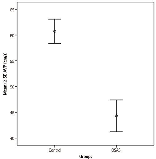
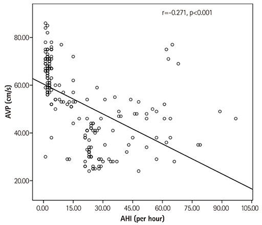
AVP and FMD were significantly decreased in patients with OSAS compared to controls (p<0.001). PWV and CIMT were increased in the OSAS group compared to controls (p<0.001). Moreover, AVP and FMD were significantly decreased in the severe OSAS group compared to the mild to moderate OSAS group (p<0.001). PWV and CIMT were significantly increased in the severe group compared to the mild to moderate group (p<0.001). AVP was significantly positively correlated with FMD (r=0.564, p<0.001). However, it was found to be significantly inversely related to PWV (r=-0.580, p<0.001) and CIMT (r=-0.251, p<0.001).
CONCLUSION
The measurement of AVP is a novel and practical echocardiographic method, which may be used to identify arterial stiffness in OSAS.
Keyword
MeSH Terms
Figure
Reference
-
1. Cavalcante JL, Lima JA, Redheuil A, Al-Mallah MH. Aortic stiffness: current understanding and future directions. J Am Coll Cardiol. 2011; 57:1511–1522.2. Çörtük M, Akyol S, Baykan AO, et al. Aortic stiffness increases in proportion to the severity of apnea-hypopnea index in patients with obstructive sleep apnea syndrome. Clin Respir J. 2014; 11. 17. [Epub]. DOI: 10.1111/crj.12244.3. Pignoli P, Tremoli E, Poli A, Oreste P, Paoletti R. Intimal plus medial thickness of the arterial wall: a direct measurement with ultrasound imaging. Circulation. 1986; 74:1399–1406.4. Ip MS, Tse HF, Lam B, Tsang KW, Lam WK. Endothelial function in obstructive sleep apnea and response to treatment. Am J Respir Crit Care Med. 2004; 169:348–353.5. Lattimore JD, Celermajer DS, Wilcox I. Obstructive sleep apnea and cardiovascular disease. J Am Coll Cardiol. 2003; 41:1429–1437.6. Drager LF, Bortolotto LA, Figueiredo AC, Silva BC, Krieger EM, Lorenzi-Filho G. Obstructive sleep apnea, hypertension, and their interaction on arterial stiffness and heart remodeling. Chest. 2007; 131:1379–1386.7. Gunes Y, Tuncer M, Guntekin U, et al. The relation between the color M-mode propagation velocity of the descending aorta and coronary and carotid atherosclerosis and flow-mediated dilatation. Echocardiography. 2010; 27:300–305.8. Simsek H, Sahin M, Gunes Y, Dogan A, Gumrukcuoglu HA, Tuncer M. A novel echocardiographic method for the detection of subclinical atherosclerosis in newly diagnosed, untreated type 2 diabetes. Echocardiography. 2013; 30:644–648.9. Simsek H, Sahin M, Gunes Y, et al. A novel echocardiographic method as an indicator of endothelial dysfunction in patients with coronary slow flow. Eur Rev Med Pharmacol Sci. 2013; 17:689–693.10. Sleep-related breathing disorders in adults: recommendations for syndrome definition and measurement techniques in clinical research. The report of an American Academy of Sleep Medicine Task Force. Sleep. 1999; 22:667–689.11. Shahar E, Whitney CW, Redline S, et al. Sleep-disordered breathing and cardiovascular disease: cross-sectional results of the Sleep Heart Health Study. Am J Respir Crit Care Med. 2001; 163:19–25.12. Wolk R, Kara T, Somers VK. Sleep-disordered breathing and cardiovascular disease. Circulation. 2003; 108:9–12.13. Laurent S, Boutouyrie P, Asmar R, et al. Aortic stiffness is an independent predictor of all-cause and cardiovascular mortality in hypertensive patients. Hypertension. 2001; 37:1236–1241.14. Mattace-Raso FU, van der Cammen TJ, Hofman A, et al. Arterial stiffness and risk of coronary heart disease and stroke: the Rotterdam Study. Circulation. 2006; 113:657–663.15. Hirata K, Kawakami M, O'Rourke MF. Pulse wave analysis and pulse wave velocity: a review of blood pressure interpretation 100 years after Korotkov. Circ J. 2006; 70:1231–1239.16. Kals J, Lieberg J, Kampus P, Zagura M, Eha J, Zilmer M. Prognostic impact of arterial stiffness in patients with symptomatic peripheral arterial disease. Eur J Vasc Endovasc Surg. 2014; 48:308–315.17. Lorenz MW, Markus HS, Bots ML, Rosvall M, Sitzer M. Prediction of clinical cardiovascular events with carotid intima-media thickness: a systematic review and meta-analysis. Circulation. 2007; 115:459–467.18. Silvestrini M, Rizzato B, Placidi F, Baruffaldi R, Bianconi A, Diomedi M. Carotid artery wall thickness in patients with obstructive sleep apnea syndrome. Stroke. 2002; 33:1782–1785.19. Schulz R, Seeger W, Fegbeutel C, et al. Changes in extracranial arteries in obstructive sleep apnoea. Eur Respir J. 2005; 25:69–74.20. Gullu H, Erdogan D, Caliskan M, et al. Interrelationship between noninvasive predictors of atherosclerosis: transthoracic coronary flow reserve, flow-mediated dilation, carotid intima-media thickness, aortic stiffness, aortic distensibility, elastic modulus, and brachial artery diameter. Echocardiography. 2006; 23:835–842.21. Tavil Y, Kanbay A, Sen N, et al. The relationship between aortic stiffness and cardiac function in patients with obstructive sleep apnea, independently from systemic hypertension. J Am Soc Echocardiogr. 2007; 20:366–372.22. Drager LF, Bortolotto LA, Lorenzi MC, Figueiredo AC, Krieger EM, Lorenzi-Filho G. Early signs of atherosclerosis in obstructive sleep apnea. Am J Respir Crit Care Med. 2005; 172:613–618.23. Wilkinson IB, McEniery CM. Arterial stiffness, endothelial function and novel pharmacological approaches. Clin Exp Pharmacol Physiol. 2004; 31:795–799.24. Shandas R, Weinberg C, Ivy DD, et al. Development of a noninvasive ultrasound color M-mode means of estimating pulmonary vascular resistance in pediatric pulmonary hypertension: mathematical analysis, in vitro validation, and preliminary clinical studies. Circulation. 2001; 104:908–913.25. Paule WJ, Zemplenyi TK, Rounds DE, Blankenhorn DH. Light- and electron-microscopic characteristics of artrial smooth muscle cell cultures subjected to hypoxia or carbon monoxide. Atherosclerosis. 1976; 25:111–123.26. Güneş Y, Tuncer M, Yildirim M, Güntekin U, Gümrükçüoğlu HA, Sahin M. A novel echocardiographic method for the prediction of coronary artery disease. Med Sci Monit. 2008; 14:MT42–MT46.27. Nichols WW. Clinical measurement of arterial stiffness obtained from noninvasive pressure waveforms. Am J Hypertens. 2005; 18(1 Pt 2):3S–10S.28. Güneş Y, Gümrükçüoğlu HA, Kaya Y, Tuncer M. Incremental diagnostic value of color M-mode propagation velocity of the descending thoracic aorta to exercise electrocardiography. Turk Kardiyol Dern Ars. 2010; 38:551–557.29. Guntekin U, Gunes Y, Gunes A, et al. Noninvasive assessment of atherosclerosis in patients with isolated hypertension. Echocardiography. 2010; 27:155–160.30. Sahin M, Simsek H, Akyol A, et al. A new echocardiographic parameter of arterial stiffness in end-stage renal disease. Herz. 2014; 39:749–754.
- Full Text Links
- Actions
-
Cited
- CITED
-
- Close
- Share
- Similar articles
-
- The Role of Endothelin-1 in Obstructive Sleep Apnea Syndrome and Pulmonary Hypertension
- A Sleepy Man with Chronic Obstructive Pulmonary Disease-Obstructive Sleep Apnea Overlap Syndrome
- Pathophysiology and Diagnosis of Sleep Apnea
- A Case of REM-Dependent Obstructive Sleep Apnea Syndrome
- Pediatric sleep questionnaires for screening of obstructive sleep apnea syndrome


