J Rheum Dis.
2015 Dec;22(6):395-400. 10.4078/jrd.2015.22.6.395.
Mikulicz's Disease with Progressively Transformed Germinal Centers-type Immunoglobulin G4-related Lymphadenopathy Mimicking Sjogren's Syndrome
- Affiliations
-
- 1Department of Internal Medicine, The Catholic University of Korea, Catholic Medical Center, Seoul, Korea.
- 2Department of Hospital Pathology, The Catholic University of Korea, Bucheon St. Mary's Hospital, Bucheon, Korea.
- 3Division of Rheumatology, Department of Internal Medicine, The Catholic University of Korea, Bucheon St. Mary's Hospital, Bucheon, Korea. rmin6403@hanmail.net
- KMID: 2222816
- DOI: http://doi.org/10.4078/jrd.2015.22.6.395
Abstract
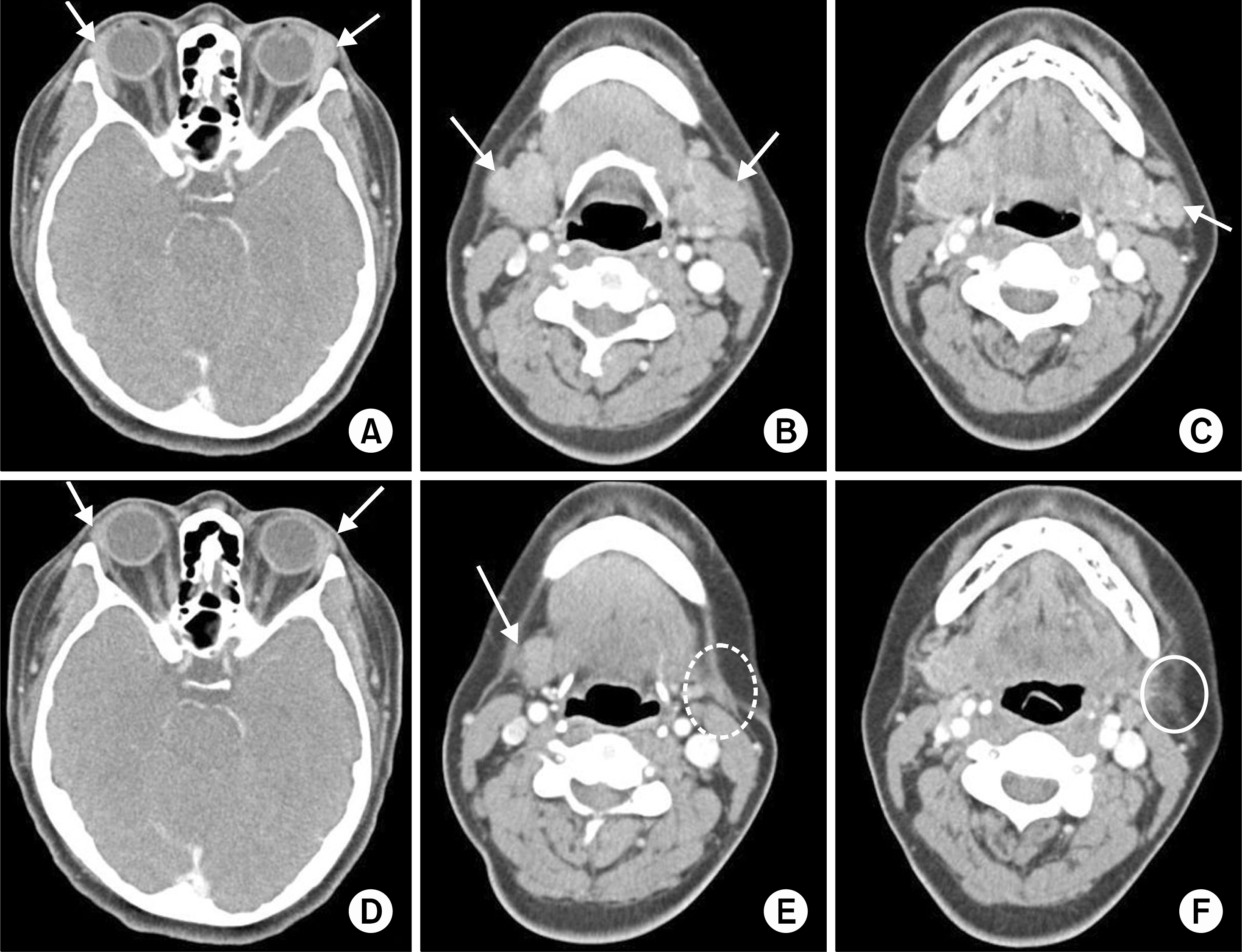
- Immunoglobulin G4-related disease (IgG4-RD) is a systemic disease, and lymphadenopathy is frequently observed in these patients. Among the 5 subtypes of IgG4-related lymphadenopathy, progressively transformed germinal centers (PTGC)-type IgG4-related lymphadenopathy possesses a unique characteristic that differentiates it from the other 4 subtypes. Here, we report on a rare case of PTGC-type IgG4-related lymphadenopathy accompanying Mikulicz's disease. A 39-year-old female complained of a left cervical mass and bilateral upper eyelid hypertrophy. The serum level of IgG4 was elevated, and computed tomography showed enlargement of the bilateral lacrimal and submandibular glands and left cervical lymph node. Excisional biopsy of a submandibular gland and cervical lymph node was performed, and the histopathologic findings revealed Mikulicz's disease accompanied by PTGC-type IgG4-related lymphadenopathy. After treatment of the patient with oral prednisolone and azathioprine, the patient's appearance improved. To the best of our knowledge, no case of PTGC-type IgG4-related lymphadenopathy has been previously reported in Korea.
Keyword
MeSH Terms
Figure
Reference
-
1. Stone JH, Zen Y, Deshpande V. IgG4-related disease. N Engl J Med. 2012; 366:539–51.
Article2. Yamamoto M, Takahashi H, Shinomura Y. Mechanisms and assessment of IgG4-related disease: lessons for the rheumatologist. Nat Rev Rheumatol. 2014; 10:148–59.
Article3. Mahajan VS, Mattoo H, Deshpande V, Pillai SS, Stone JH. IgG4-related disease. Annu Rev Pathol. 2014; 9:315–47.
Article4. Sato Y, Yoshino T. IgG4-related lymphadenopathy. Int J Rheumatol. 2012; 2012; 572539.
Article5. Yao Q, Wu G, Hoschar A. IgG4-related Mikulicz's disease is a multiorgan lymphoproliferative disease distinct from Sjögren's syndrome: a Caucasian patient and literature review. Clin Exp Rheumatol. 2013; 31:289–94.6. Ferry JA. IgG4-related lymphadenopathy and IgG4-related lymphoma: moving targets. Diagn Histopathol. 2013; 19:128–39.
Article7. Jones D. Dismantling the germinal center: comparing the processes of transformation, regression, and fragmentation of the lymphoid follicle. Adv Anat Pathol. 2002; 9:129–38.
Article8. Nguyen PL, Ferry JA, Harris NL. Progressive transformation of germinal centers and nodular lymphocyte predominance Hodgkin's disease: a comparative immunohistochemical study. Am J Surg Pathol. 1999; 23:27–33.9. Sato Y, Inoue D, Asano N, Takata K, Asaoku H, Maeda Y, et al. Association between IgG4-related disease and progressively transformed germinal centers of lymph nodes. Mod Pathol. 2012; 25:956–67.
Article10. Takeuchi M, Sato Y, Ohno K, Tanaka S, Takata K, Gion Y, et al. T helper 2 and regulatory T-cell cytokine production by mast cells: a key factor in the pathogenesis of IgG4-related disease. Mod Pathol. 2014; 27:1126–36.
Article11. Cheuk W, Chan JK. Lymphadenopathy of IgG4-related disease: an underdiagnosed and overdiagnosed entity. Semin Diagn Pathol. 2012; 29:226–34.
Article12. Umehara H, Okazaki K, Masaki Y, Kawano M, Yamamoto M, Saeki T, et al. Comprehensive diagnostic criteria for IgG4-related disease (IgG4-RD), 2011. Mod Rheumatol. 2012; 22:21–30.
Article13. Khosroshahi A, Stone JH. Treatment approaches to IgG4-related systemic disease. Curr Opin Rheumatol. 2011; 23:67–71.
Article14. Hart PA, Topazian MD, Witzig TE, Clain JE, Gleeson FC, Klebig RR, et al. Treatment of relapsing autoimmune pancreatitis with immunomodulators and rituximab: the Mayo Clinic experience. Gut. 2013; 62:1607–15.
Article
- Full Text Links
- Actions
-
Cited
- CITED
-
- Close
- Share
- Similar articles
-
- A Case of Immunoglobulin G4-Related Sialadenitis and Dacryoadenitis
- Unusual Multiorgan Immunoglobulin G4 (IgG4) Inflammation: Autoimmune Pancreatitis, Mikulicz Syndrome, and IgG4 Mastitis
- Overview of the Immunoglobulin G4-related Disease Spectrum
- Poor positive predictive value of serum immunoglobulin G4 concentrations in the diagnosis of immunoglobulin G4-related sclerosing disease
- Update of Sjogren's Syndrome


