
Clinical Implication of 18F-FDG-PET in Diagnosing and Monitoring Disease Activity in a Case of Subclinical Stage of Giant Cell Arteritis
- Affiliations
-
- 1Department of Internal Medicine, Chungnam National University School of Medicine, Daejeon, Korea. kangsw@cnuh.co.kr
- 2Department of Nuclear Medicine, Chungnam National University School of Medicine, Daejeon, Korea.
- KMID: 2222813
- DOI: http://doi.org/10.4078/jrd.2015.22.6.382
Abstract
- Giant cell arteritis (GCA) is a systemic vasculitis which typically occurs in persons over 50 years old. GCA is closely related to polymyalgia rheumatica (PMR). A temporal artery biopsy is the gold standard test for the diagnosis of GCA. Recently, there is increasing evidence for the role of 18F-fluorodeoxyglucose positron emission tomography (18F-FDG-PET) in diagnosis of vasculitis. Here, we report on a case of a 67-year-old Korean male who was diagnosed with atypical GCA in subclinical stage concomitant with PMR by 18F-FDG-PET. After treatment, abnormal findings of 18F-FDG-PET were improved.
MeSH Terms
Figure
-
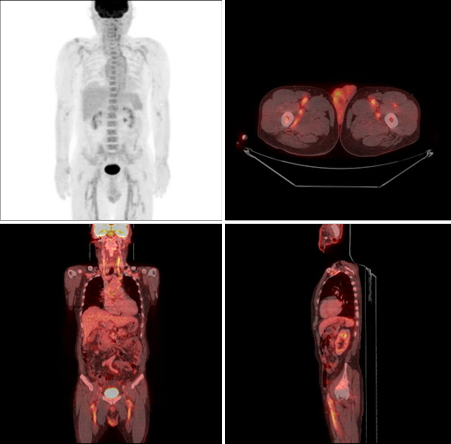
Figure 1. 18 F-fluorodeoxyglucose positron emission tomography before treatment. There are increased glucose metabolism in the walls of the ascending aorta, aortic arch, thoracic descending aorta, both subclavian arteries, both common carotid arteries and especially both common femoral arteries and their large branches with wall thickening.
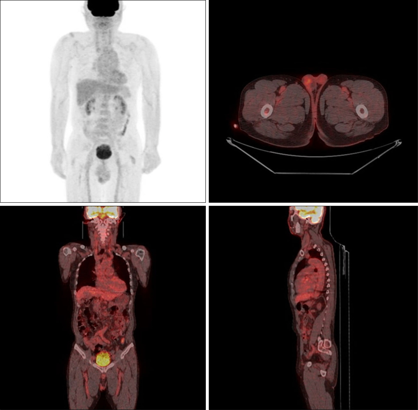
Figure 2. 18 F-fluorodeoxyglucose positron emission tomography after 12 months of treatment. There is no metabolic evidence of arteritis.
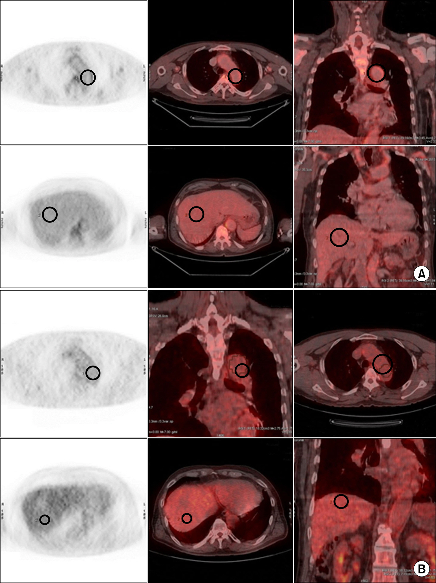
Figure 3. 18 F-fluorodeoxyglucose positron emission tomography images and aorta to liver maximal standardized uptake value ratio. (A) The aorta to liver ratio before treatment was 1.49 (thoracic aorta 3.45, liver 2.32). (B) The aorta to liver ratio after treatment was 0.98 (thoracic aorta 2.75, liver 2.80).
Reference
-
1. Patil P, Karia N, Jain S, Dasgupta B. Giant cell arteritis: a review. Eye Brain. 2013; 5:23–33.2. Firestein GS, Budd RC, Gabriel SE, McInnes IB, O'Dell JR. Kelley's textbook of rheumatology. 9th ed.Philadelphia, PA: Saunders;2013. p. 1461–5.3. Salvarani C, Cantini F, Boiardi L, Hunder GG. Polymyalgia rheumatica and giant-cell arteritis. N Engl J Med. 2002; 347:261–71.
Article4. Hautzel H, Sander O, Heinzel A, Schneider M, Müller HW. Assessment of large-vessel involvement in giant cell arteritis with 18F-FDG PET: introducing an ROC-analysis-based cutoff ratio. J Nucl Med. 2008; 49:1107–13.
Article5. Kermani TA, Warrington KJ. Polymyalgia rheumatica. Lancet. 2013; 381:63–72.
Article6. Salvarani C, Gabriel SE, O'Fallon WM, Hunder GG. Epidemiology of polymyalgia rheumatica in Olmsted County, Minnesota, 1970-1991. Arthritis Rheum. 1995; 38:369–73.
Article7. Gonzalez-Gay MA, Garcia-Porrua C, Llorca J, Gonzalez-Louzao C, Rodriguez-Ledo P. Biopsy-negative giant cell arteritis: clinical spectrum and predictive factors for positive temporal artery biopsy. Semin Arthritis Rheum. 2001; 30:249–56.
Article8. Roth AM, Milsow L, Keltner JL. The ultimate diagnoses of patients undergoing temporal artery biopsies. Arch Ophthalmol. 1984; 102:901–3.
Article9. Fletcher TM, Espinola D. Positron emission tomography in the diagnosis of giant cell arteritis. Clin Nucl Med. 2004; 29:617–9.
Article10. Besson FL, Parienti JJ, Bienvenu B, Prior JO, Costo S, Bouvard G, et al. Diagnostic performance of 18 F-fluorodeox-yglucose positron emission tomography in giant cell arteritis: a systematic review and meta-analysis. Eur J Nucl Med Mol Imaging. 2011; 38:1764–72.11. Kang MI, Park HJ, Jeon HM, Kang Y, Lee SW, Lee SK, et al. A case of atypical giant cell arteritis presenting as a fever of unknown origin. J Rheum Dis. 2012; 19:299–301.
Article12. Kwon CM, Hong YH, Chun KA, Cho IH, Kim MJ, Shin DG, et al. A case of silent giant cell arteritis involving the entire aorta, carotid artery and brachial artery screened by integrated PET/CT. Clin Rheumatol. 2007; 26:1959–62.
Article13. Dasgupta B, Cimmino MA, Kremers HM, Schmidt WA, Schirmer M, Salvarani C, et al. 2012 provisional classification criteria for polymyalgia rheumatica: a European League Against Rheumatism/American College of Rheumatology collaborative initiative. Arthritis Rheum. 2012; 64:943–54.
Article14. Hunder GG, Bloch DA, Michel BA, Stevens MB, Arend WP, Calabrese LH, et al. The American College of Rheumatology 1990 criteria for the classification of giant cell arteritis. Arthritis Rheum. 1990; 33:1122–8.
Article15. Camellino D, Cimmino MA. Imaging of polymyalgia rheumatica: indications on its pathogenesis, diagnosis and prognosis. Rheumatology (Oxford). 2012; 51:77–86.
Article
- Full Text Links
-
- Actions
-
Cited
- CITED
-
- Close
- Share
-
- Similar articles
-
- A Case of Atypical Giant Cell Arteritis Presenting as a Fever of Unknown Origin
- Application of Simultaneous 18F-FDG PET/MRI for Evaluating Residual Lesion in Pyogenic Spine Infection:a Case Report
- FDG PET-CT in the Diagnosis of Takayasu Arteritis Presenting as Fever of Unknown Origin: A Case Report
- Giant Cell Tumor Pulmonary Metastases Mimic Primary Malignant Pulmonary Nodules on 18F-FDG PET/CT
- Giant cell arteritis of the breast: a case report


