A Case of Epstein-Barr Virus-associated Hydroa Vacciniforme
- Affiliations
-
- 1Department of Dermatology, Seoul National University College of Medicine, Seoul, Korea. khcho@snu.ac.kr
- KMID: 2219400
- DOI: http://doi.org/10.5021/ad.2009.21.2.209
Abstract
- Hydroa vacciniforme (HV) is a photosensitivity disorder characterized by recurrent necrotic vesiculopapules on sun-exposed areas, which heal spontaneously during adolescence. Recently, an association has been reported between latent Epstein-Barr virus (EBV) infection and atypical HV-like eruption and malignant potential. However, latent EBV infection has also been reported in the setting of typical HV. An 11-year-old girl presented with recurrent, scattered, discrete vesicular eruptions with scarring on the face and the extensor surfaces of both forearms. In-situ hybridization was carried out to detect latent EBV infection. Based on the clinical and histopathological findings, typical EBV-associated HV was suspected.
Keyword
MeSH Terms
Figure
-
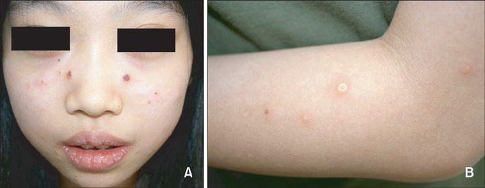
Fig. 1 The patient showed multiple, discrete vesicular eruptions with scarring on the face (A) and the extensor surfaces of the forearms (B).
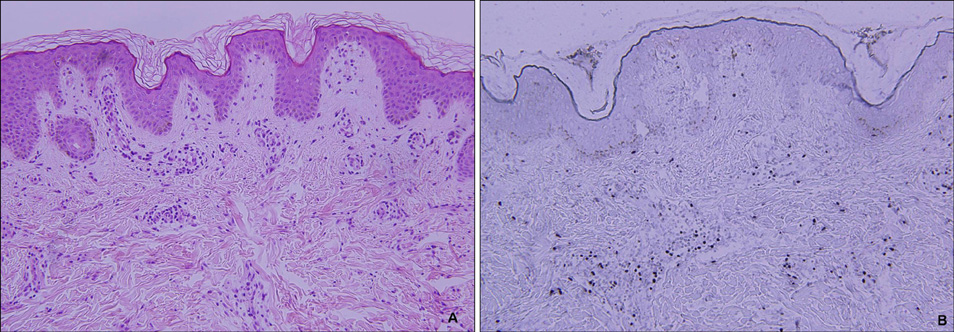
Fig. 2 (A) A skin biopsy obtained from the vesicular lesion on her forearm. Intraepidermal vesiculation with superficial and deep perivascular and periappendageal infiltrations in the upper dermis (H&E, ×200). (B) In situ hybridization. Many cells positive for EBV-encoded small nuclear RNA are present in the infiltrates (×200).
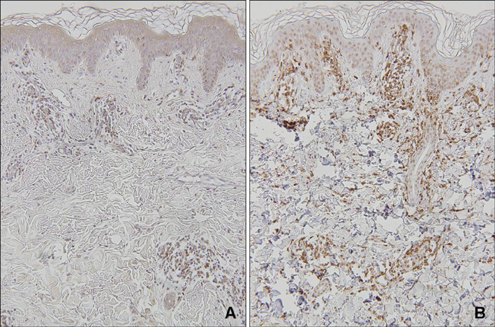
Fig. 3 Immunohistochemical stains. The dermal infiltrates are composed mainly of lymphocytes expressing CD3-positive (A) and CD45RO-positive (B) cells (Immunoperoxidase stain, ×100).
Cited by 1 articles
-
Epstein-Barr Virus-Associated Vesiculopapular Eruption on the Face of a Patient with Natural Killer T Cell Lymphoma
Ji Soo Lim, Tae Min Kim, Kwang Hyun Cho
Ann Dermatol. 2017;29(5):618-620. doi: 10.5021/ad.2017.29.5.618.
Reference
-
1. Cho KH, Kim CW, Lee DY, Sohn SJ, Kim DW, Chung JH. An Epstein-Barr virus-associated lymphoproliferative lesion of the skin presenting as recurrent necrotic papulovesicles of the face. Br J Dermatol. 1996. 134:791–796.
Article2. Cho KH, Kim CW, Kwon OS, Yang SG, Park KC, Park MH, et al. Epstein-Barr virus-associated lymphoproliferative eruption with progression to large granular lymphocytic leukaemia. Br J Dermatol. 1997. 137:426–430.
Article3. Cho KH, Kim CW, Heo DS, Lee DS, Choi WW, Rim JH, et al. Epstein-Barr virus-associated peripheral T-cell lymphoma in adults with hydroa vacciniforme-like lesions. Clin Exp Dermatol. 2001. 26:242–247.
Article4. Magana M, Sangueza P, Gil-Beristain J, Sanchez-Sosa S, Salgado A, Ramon G, et al. Angiocentric cutaneous T-cell lymphoma of childhood (hydroa-like lymphoma): a distinctive type of cutaneous T-cell lymphoma. J Am Acad Dermatol. 1998. 38:574–579.
Article5. Ruiz-Maldonado R, Parrilla FM, Orozco-Covarrubias ML, Ridaura C, Tamayo Sanchez L, Duran McKinster C. Edematous, scarring vasculitic panniculitis: a new multisystemic disease with malignant potential. J Am Acad Dermatol. 1995. 32:37–44.
Article6. Oono T, Arata J, Masuda T, Ohtsuki Y. Coexistence of hydroa vacciniforme and malignant lymphoma. Arch Dermatol. 1986. 122:1306–1309.
Article7. Asada H, Okada N, Tei H, Yamamura T, Hashimoto K, Kondo K, et al. Epstein-Barr virus-associated large granular lymphocyte leukemia with cutaneous infiltration. J Am Acad Dermatol. 1994. 31:251–255.
Article8. Iwatsuki K, Xu Z, Takata M, Iguchi M, Ohtsuka M, Akiba H, et al. The association of latent Epstein-Barr virus infection with hydroa vacciniforme. Br J Dermatol. 1999. 140:715–721.
Article9. Hann SK, Im S, Park YK, Lee S. Hydroa vacciniforme with unusually severe scar formation: diagnosis by repetitive UVA phototesting. J Am Acad Dermatol. 1991. 25:401–403.
Article10. Paik SA, Hahm JH, Kook HI. Three cases of hydroa vacciniforme. Korean J Dermatol. 1977. 15:341–345.11. Lim SD. A case of hydroa vacciniforme. Korean J Dermatol. 1960. 1:65–67.12. Chang SH, Yoon TY. A case of hydroa vacciniforme with ocular involvment. Korean J Dermatol. 1993. 31:612–615.13. Bazin E, Baudot E, Guérard L. Leçons théoriques et cliniques sur les affections génériques de la peau. 1862. Vol. 1. Paris: Delahaye;132.14. Bickers DR, Demar LK, DeLeo V, Poh-Fitzpatrick MB, Aronberg JM, Harber LC. Hydroa vacciniforme. Arch Dermatol. 1978. 114:1193–1196.
Article15. Iwatsuki K, Satoh M, Yamamoto T, Oono T, Morizane S, Ohtsuka M, et al. Pathogenic link between hydroa vacciniforme and Epstein-Barr virus-associated hematologic disorders. Arch Dermatol. 2006. 142:587–595.
Article16. Iwatsuki K, Ohtsuka M, Harada H, Han G, Kaneko F. Clinicopathologic manifestations of Epstein-Barr virus-associated cutaneous lymphoproliferative disorders. Arch Dermatol. 1997. 133:1081–1086.
Article17. Wu YH, Chen HC, Hsiao PF, Tu MI, Lin YC, Wang TY. Hydroa vacciniforme-like Epstein-Barr virus-associated monoclonal T-lymphoproliferative disorder in a child. Int J Dermatol. 2007. 46:1081–1086.
Article18. Iwatsuki K, Xu Z, Ohtsuka M, Kaneko F. Cutaneous lymphoproliferative disorders associated with Epstein-Barr virus infection: a clinical overview. J Dermatol Sci. 2000. 22:181–195.
Article19. Cho KH, Lee SH, Kim CW, Jeon YK, Kwon IH, Cho YJ, et al. Epstein-Barr virus-associated lymphoproliferative lesions presenting as a hydroa vacciniforme-like eruption: an analysis of six cases. Br J Dermatol. 2004. 151:372–380.
Article20. Rowe M, Lear AL, Croom-Carter D, Davies AH, Rickinson AB. Three pathways of Epstein-Barr virus gene activation from EBNA1-positive latency in B lymphocytes. J Virol. 1992. 66:122–131.
Article
- Full Text Links
-
- Actions
-
Cited
- CITED
-
- Close
- Share
-
- Similar articles
-
- Epstein Barr Virus-associated Vesicular Eruptions of the Face
- Epstein-Barr Virus Associated Hydroa Vacciniforme-like Eruption
- Hydroa Vacciniforme:Diagnosis by Repetitive UVA Phototesting
- Epstein-Barr Virus-Associated Vesiculopapular Eruption on the Face of a Patient with Natural Killer T Cell Lymphoma
- A case of hydroa vacciniforme with ocular involvment
