Ann Dermatol.
2009 May;21(2):154-158. 10.5021/ad.2009.21.2.154.
Osteoma Cutis as the Presenting Feature of Albright Hereditary Osteodystrophy Associated with Pseudopseudohypoparathyroidism
- Affiliations
-
- 1Department of Dermatology, College of Medicine, Kyunghee University, Seoul, Korea. bellotte@hanmail.net
- KMID: 2219385
- DOI: http://doi.org/10.5021/ad.2009.21.2.154
Abstract
- Primary osteoma cutis is characterized by the formation of normal bone tissue in the dermis or subcutis without any underlying tissue abnormality or pre-existing calcification. This illness is associated with Albright hereditary osteodystrophy (AHO), which is characterized by such physical features as a short stature, round face, obesity, brachydactyly and osteoma cutis. Pseudohypoparathyroidism (PHP) is an inherited metabolic disorder that's characterized by resistance to parathyroid hormone, and PHP is present in most AHO patients. An AHO phenotype without hormonal resistance is called pseudopseudohypoparathyroidism (PPHP). Osteoma cutis is less common in patients with PPHP than in patients with PHP. We present here a case of osteoma cutis as the cardinal manifestation of AHO associated with PPHP. Osteoma cutis is an important sign of AHO and its significance should not be overlooked, even if the patient has normal values on the serum biochemical tests.
Keyword
MeSH Terms
-
Alkenes
Bone and Bones
Bone Diseases, Metabolic
Brachydactyly
Dermis
Fibrous Dysplasia, Polyostotic
Humans
Obesity
Ossification, Heterotopic
Osteoma
Parathyroid Hormone
Phenotype
Pseudohypoparathyroidism
Pseudopseudohypoparathyroidism
Reference Values
Skin Diseases, Genetic
Alkenes
Bone Diseases, Metabolic
Fibrous Dysplasia, Polyostotic
Ossification, Heterotopic
Parathyroid Hormone
Skin Diseases, Genetic
Figure
-
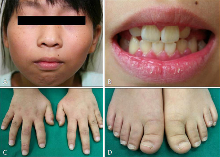
Fig. 1 Photograph showing the round face (A), malocclusion of teeth (B), the shortened 4th and 5th fingers (C) and the shortened left 4th toe (D).
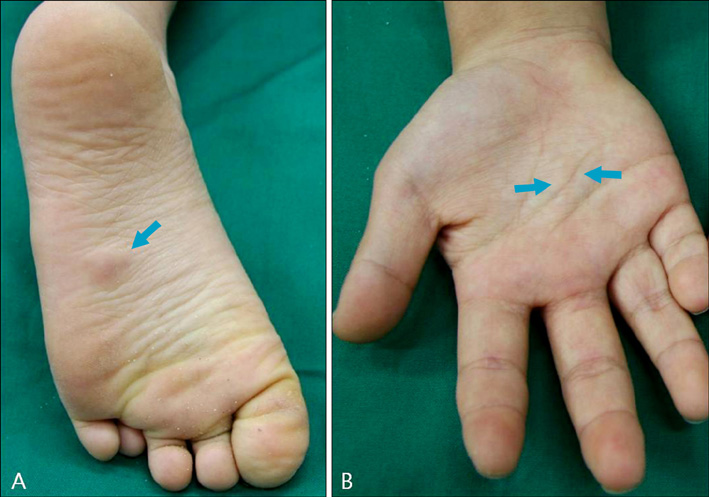
Fig. 2 Presence of skin-colored, hard nodules on the left sole (A) and right palm (B).
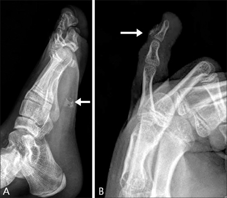
Fig. 3 Extraskeletal ossifications are seen on the left sole (A) and left 5th finger (B).
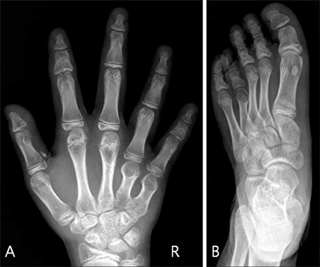
Fig. 4 Shortened right and left 4th and 5th metacarpal bones (A) and the shortened left 4th metatarsal bone (B).

Fig. 5 Small spicules to large masses of mature bone are found in the dermis (A: Lt. 5th finger, B: Lt. sole, ×40). The bony tissue is composed of osteocytes, osteoblasts, Haversian canals and condensed mesenchyme (C: Lt. sole, ×100).
Reference
-
1. Albright F, Burnett CH, Smith PH, Parson W. Pseudohypoparathyroidism: an example of Seabright-Bantam syndrome. Endocrinology. 1942. 30:922–932.2. Novak C, Siller G, Wood D. Idiopathic multiple miliary osteomas of the face. Australas J Dermatol. 1998. 39:109–111.
Article3. Roth SI, Stowell RE, Helwig EB. Cutaneous ossification. Report of 120 cases and review of the literature. Arch Pathol. 1963. 76:44–54.4. Sethuraman G, Malhotra AK, Khaitan BK, Kumar R, Sharma VK, Kabra M, et al. Osteoma cutis in pseudohypoparathyroidism. Clin Exp Dermatol. 2006. 31:225–227.
Article5. Walsh JS, Fairley JA. Wolff K, Goldsmith LA, Katz SI, Gilchrest BA, Paller AS, Leffell DJ, editors. Cutaneous mineralization and ossification. Fitzpatrick's dermatology in general medicine. 2008. 7th ed. New York: McGraw-Hill;1296–1297.6. Kapoor S, Gogia S, Paul R, Banerjee S. Albright's hereditary osteodystrophy. Indian J Pediatr. 2006. 73:153–156.
Article7. Trueb RM, Panizzon RG, Burg G. Cutaneous ossification in Albright's hereditary osteodystrophy. Dermatology. 1993. 186:205–209.
Article8. Wilson LC, Hall CM. Albright's hereditary osteodystrophy and pseudohypoparathyroidism. Semin Musculoskelet Radiol. 2002. 6:273–283.
Article9. Gelfand IM, Hub RS, Shore EM, Kaplan FS, Dimeglio LA. Progressive osseous heteroplasia-like heterotopic ossification in a male infant with pseudohypoparathyroidism type Ia: a case report. Bone. 2007. 40:1425–1428.
Article10. Lietman SA, Ding C, Cooke DW, Levine MA. Reduction in Gsalpha induces osteogenic differentiation in human mesenchymal stem cells. Clin Orthop Relat Res. 2005. 231–238.11. Long DN, McGuire S, Levine MA, Weinstein LS, Germain-Lee EL. Body mass index differences in pseudohypoparathyroidism type 1a versus pseudopseudohypoparathyroidism may implicate paternal imprinting of Galpha(s) in the development of human obesity. J Clin Endocrinol Metab. 2007. 92:1073–1079.
Article12. Eyre WG, Reed WB. Albright's hereditary osteodystrophy with cutaneous bone formation. Arch Dermatol. 1971. 104:634–642.
Article13. Prendiville JS, Lucky AW, Mallory SB, Mughal Z, Mimouni F, Langman CB. Osteoma cutis as a presenting sign of pseudohypoparathyroidism. Pediatr Dermatol. 1992. 9:11–18.
Article14. Monn E, Osnes JB, Oye I, Wefring KW. Pseudohypoparathyroidism: a difficult diagnosis in early childhood. Acta Paediatr Scand. 1976. 65:487–493.15. Burnstein MI, Kottamasu SR, Pettifor JM, Sochett E, Ellis BI, Frame B. Metabolic bone disease in pseudohypoparathyroidism: radiologic features. Radiology. 1985. 155:351–356.
Article
- Full Text Links
-
- Actions
-
Cited
- CITED
-
- Close
- Share
-
- Similar articles
-
- Osteoma cutis in Albright's Hereditary Osteodystrophy
- A Case of Osteoma Cutis, a Diagnostic Clue for Albright's Hereditary Osteodystrophy
- A Case of Primary Osteoma Cutis
- Two Cases of Albright's Hereditary Osteodystrophy Occurring in Pseudopseudohypoparathyroidism
- A Case of Multiple Osteoma Cutis in Infant
