J Korean Ophthalmol Soc.
2013 Jun;54(6):962-965. 10.3341/jkos.2013.54.6.962.
A Case of Idiopathic Orbital Sudoriferous Cyst in an Adult
- Affiliations
-
- 1Department of Ophthalmology, Chonbuk National University Medical School, Research Institute of Clinical Medicine of Chonbuk National University-Biomedical Research Institute of Chonbuk National University Hospital, Jeonju, Korea. ahnmin@jbnu.ac.kr
- KMID: 2217275
- DOI: http://doi.org/10.3341/jkos.2013.54.6.962
Abstract
- PURPOSE
To report a rare case of an idiopathic sudoriferous cyst involving the orbit in an adult.
CASE SUMMARY
A 52-year-old male visited our clinic with an orbital mass in the right eye, which had developed 4 months prior to admission. A 2 x 1.5 cm-sized hard orbital mass was palpated in the middle area of the right upper orbit. On sonographic imaging, a well-demarcated tumor was identified that showed no echogenicity. We performed anterior orbitotomy with a lid crease incision. The tumor was completely removed. Histopathological examination showed a solitary cyst lined with two or three layers of cuboidal epithelial cells with scattered goblet cells. The tumor was classified as a sudoriferous cyst.
CONCLUSIONS
Sudoriferous cyst should be considered in the differential diagnosis of a well-circumscribed mass lesion involving the orbit in adult patients.
Keyword
Figure
-
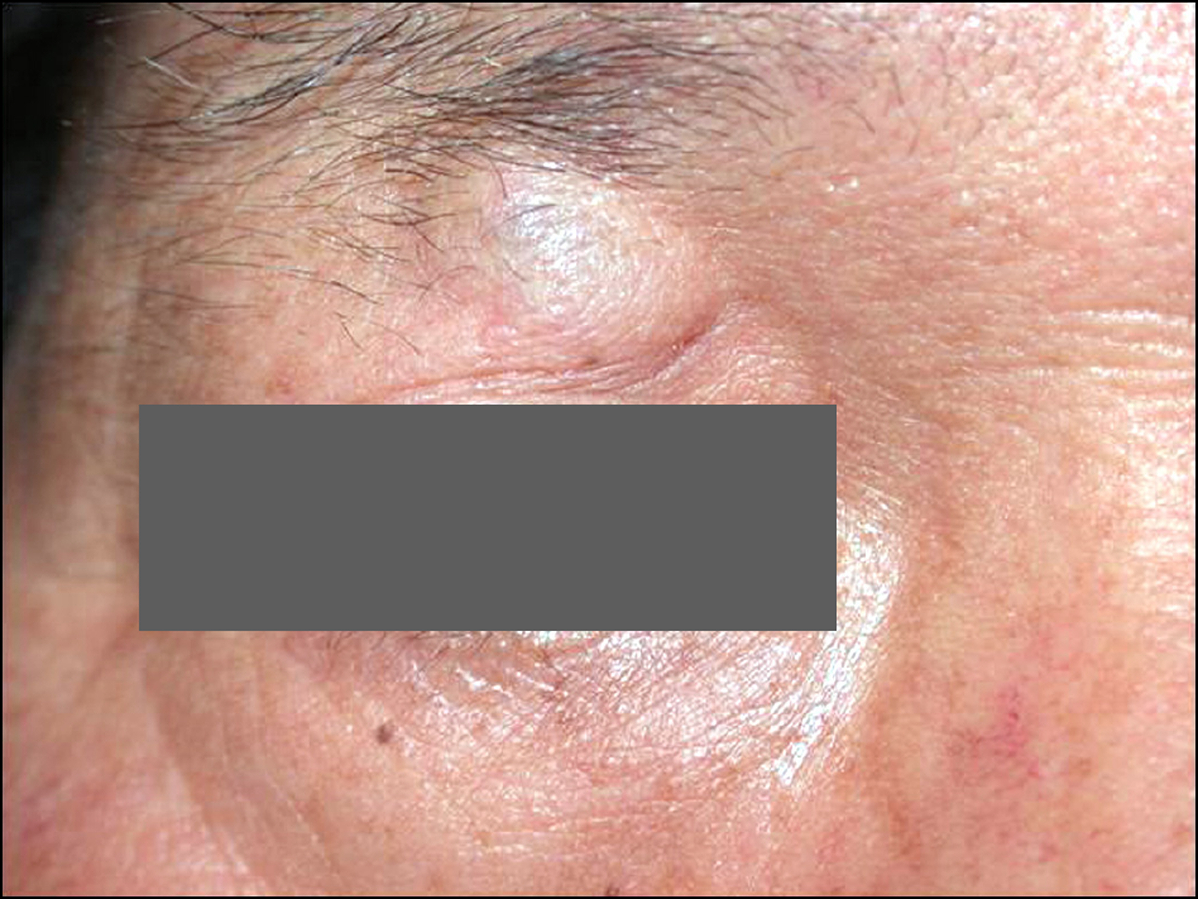
Figure 1. Preoperative photograph showing right superomedial orbital swelling, fixed to the deep orbital planes on palpation.

Figure 2. The ultrasound image of the right superomedial orbit showing the well-demarcated, thin walled, anechoic mass, consistent with cyst beneath the muscle layer.

Figure 3. Hispathologic photographs of the specimen. (A) Low power view of the cyst showing areas of ductal cyst with fibrous tis-sue wall (H&E stain, ×40). (B) High power view of the cyst showing areas of double layered cuboidal cell lining, the inner layer showing apical snouts (H&E stain, ×400). (C) High power view of the cyst showing Periodic acid Schiff (PAS) positive apical gly-cocalyx (PAS stain, ×400).
Reference
-
References
1. Wiliam HS. Eyelids and Lacrimal Drainage System. In: Jurij RB, ed. Ophthalmic Pathology. 4th ed.Philadelphia: WB Saunders;1996; v. 4:chap. 11.2. Haider E, Saigal G, Gill D. . Congenital orbital sudoriferous cyst: radiological findings. Pediatr Radiol. 2005; 35:1142–4.
Article3. Kim JY, Lee EK, Lee YH, Lee SB. Apocrine sudoriferous cyst in medial canthal region occurred after trauma. J Korean Ophthalmol Soc. 2007; 48:1562–6.
Article4. Smith RJ, Kuo IC, Reviglio VE. Multiple apocrine hidrocystomas of the eyelids. Orbit. 2012; 31:140–2.
Article5. Mehta A, Rao A, Khanna A. Sudoriferous cyst of the orbit of adult origin after trauma. Indian J Ophthalmol. 2008; 56:235–7.
Article6. Chung JK, Lee SJ, Kang SK, Park SH. Congenital sudoriferous cyst within the orbit followed by esotropia. Korean J Ophthalmol. 2007; 21:120–3.
Article7. Rosen WJ, Li Y. Sudoriferous cyst of the orbit. Ophthal Plast Reconstr Surg. 2001; 17:73–5.
Article8. Valenzuela AA, Heathcote JG. Apocrine hidrocystoma of the orbit. Orbit. 2011; 30:316–7.
Article9. Saunders JF. Congenital sudoriferous cyst of the orbit. Arch Ophthalmol. 1973; 89:205–6.
Article10. Park SJ, Jang JW, Chin HS. Sudoriferous cyst of the orbit. J Korean Ophthalmol Soc. 1998; 39:1288–90.11. Mims J, Rodrigues M, Calhoun J. Sudoriferous cyst of the orbit. Can J Ophthalmol. 1977; 12:155–6.12. Vignes JR, Franco-Vidal V, Eimer S, Liguoro D. Intraorbital apoc-rine hidrocystoma. Clin Neurol Neurosurg. 2007; 109:631–3.
Article13. Shields JA, Shields CL. Orbital cysts of childhood–classification, clinical features, and management. Surv Ophthalmol. 2004; 49:281–99.
Article
- Full Text Links
-
- Actions
-
Cited
- CITED
-
- Close
- Share
-
- Similar articles
-
- Sudoriferous Cyst of the Orbit
- Apocrine Sudoriferous Cyst in Medial Canthal Region Occurred after Trauma
- Congenital Sudoriferous Cyst within the Orbit Followed by Esotropia
- Sudoriferous Cyst Adhered to Levator Aponeurosis: A Case Report
- A Case of Idiopathic Orbital Myositis Involving All Extraocular Muscles of Both Eyes
