A Case of Pigmented Free-Floating Posterior Vitreous Cyst
- Affiliations
-
- 1Department of Ophthalmology and Visual Science, Seoul St. Mary's Hospital, The Catholic University of Korea College of Medicine, Seoul, Korea. parkyh@catholic.ac.kr
- KMID: 2217128
- DOI: http://doi.org/10.3341/jkos.2014.55.9.1392
Abstract
- PURPOSE
To report a case of a 59-year-old female with a free-floating monolateral vitreous cyst localized in the posterior vitreous in the left eye.
CASE SUMMARY
A 59-year-old female who complained of an intermittent floater in the left eye for 3 months visited our clinic. She had been suffering from visual disturbance for approximately 3 months. There was no previous history of trauma, infection, or inflammatory disorders. The best corrected visual acuity was 20/20 in both eyes. On fundoscopic exam, a 3-4 disc diameter (DD) sized, brown-colored pigmented vitreous cyst was detected at the inferior temporal side of the posterior vitreous in her left eye. B-scan ultrasound confirmed the presence of an echo-free cystic formation that was free from surrounding vitreous strands or other adhesions located at the posterior vitreous. No specific findings or leakage were observed on fluorescein angiography. We followed-up the patient periodically (1 month, 3 months, and 6 months after the initial visit) and monitored whether the size or location of the cyst had changed. At every follow-up exam, the size or location of the cyst was stationary and the patient's visual acuity was 20/20 in the affected eye, thus we suggested she should be followed-up periodically for her cyst without any intervention.
CONCLUSIONS
We report a case of a patient with no previous ocular history or impaired vision who had a free-floating vitreous cyst localized in the posterior vitreous in the left eye. The disease did not appear to progress or become aggravated over a short-term follow-up period and no specific treatment was required.
Keyword
MeSH Terms
Figure
-

Figure 1. There were fundus photographs of both eyes (A, B, C, D). Compared to the fundus photograph, which is normal, in right eye (A), a pigmented free-floating vitreous cyst (about 3-4 DD sized) at inferotemporal side in left eye was observed (B, C, D).

Figure 2. Slit-lamp photograph of the free-floating vitreus cyst. It is observed to be round, and to have a smooth surface and brown-pigments in its wall.
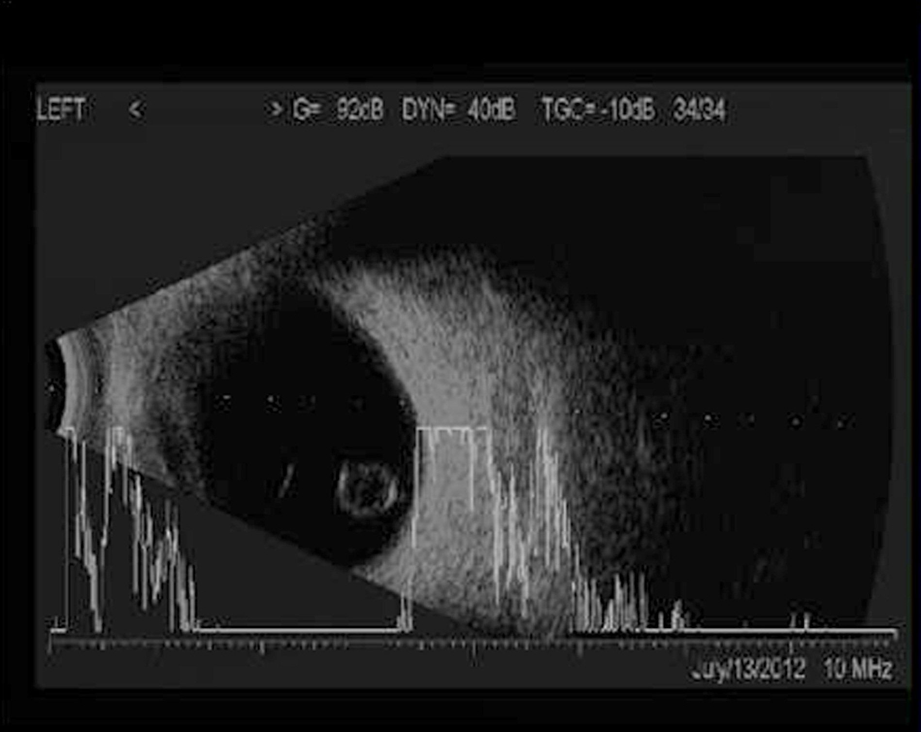
Figure 3. B-scan ultrasonography of the cyst. Note that there are no vitreous strands or adhesions.
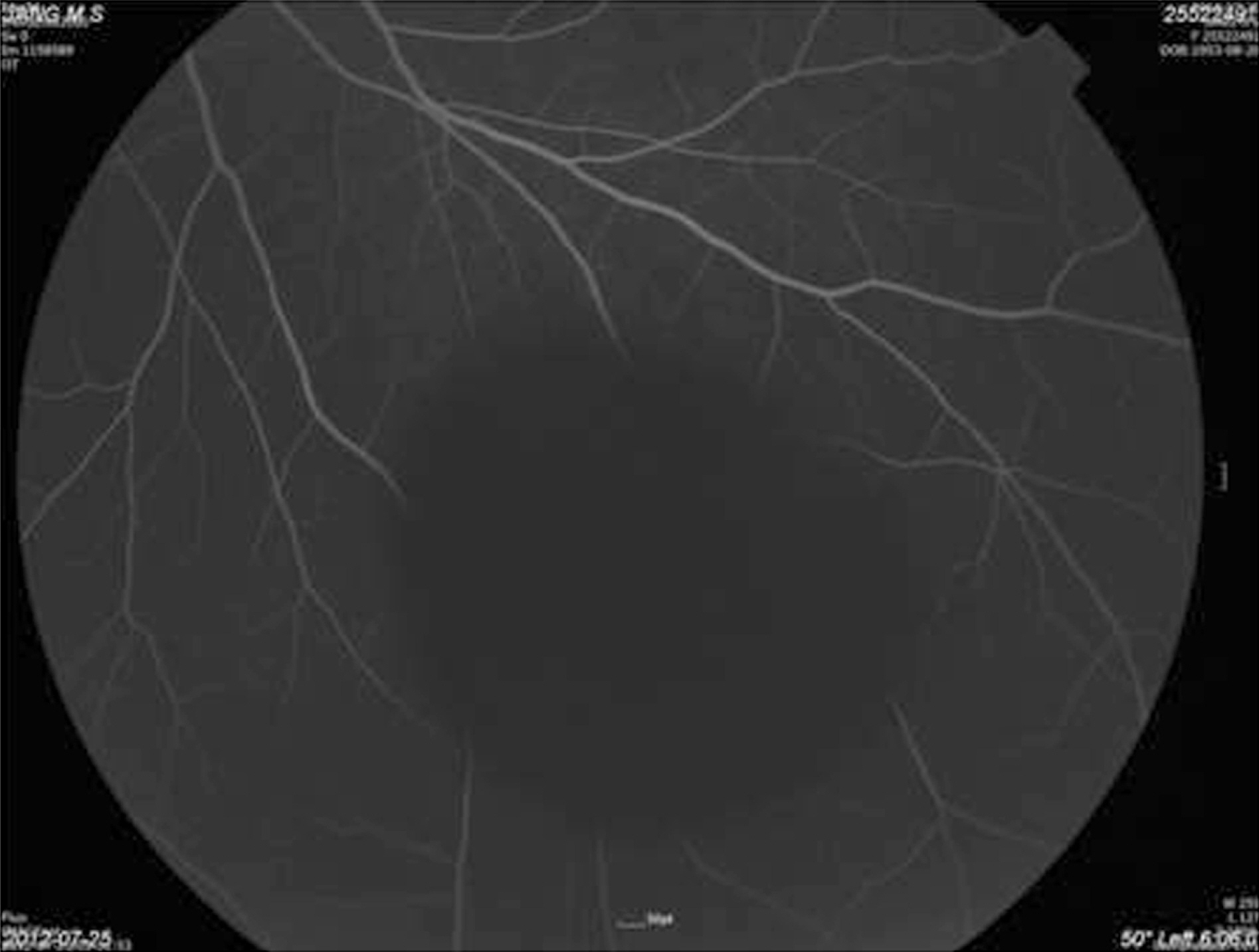
Figure 4. On fluorescein angiography, no fluorescein dye leakage either on or in the cyst were observed.
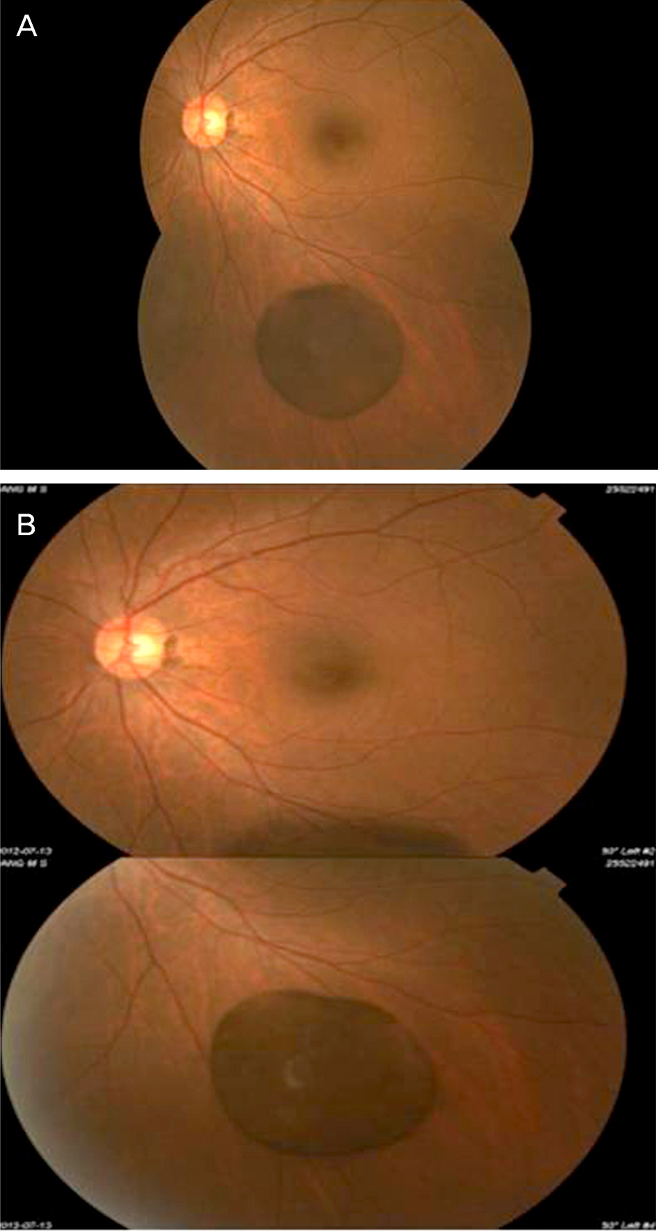
Figure 5. Fundus photographs of pigmented free-floating vitreous cyst initially (A) and after 6 months (B). Note that there was no size change or appearance (B) comparing to the first visit (A).
Reference
-
References
1. Tansley JO. Cyst of the vitreous. Trans Am Ophthalmol Soc. 1899; 8:507–9.2. Aydin E, Demir HD, Tasliyurt T. Idiopathic pigmented free-floating posterior vitreous cyst. Int Ophthalmol. 2009; 29:299–301.
Article3. Yang JE, Baek TM, Kim JH, Lee JH. A case of free-floating vitreous cyst. J Korean Ophthalmol Soc. 1990; 31:1218–20.4. Cruciani F, Santino G, Salandri AG. Monolateral idiopathic cyst of the vitreous. Acta Ophthalmol Scand. 1999; 77:601–3.
Article5. Orellana J, O'Malley RE, McPherson AR, Font RL. Pigmented free-floating vitreous cysts in two young adults. Electron microscopic observations. Ophthalmology. 1985; 92:297–302.6. Bayraktar Z, Kapran Z, Ozdogan S. Pigmented congenital vitreous cyst. Eur J Ophthalmol. 2004; 14:156–8.
Article7. Wolter JR, Martony CL, Smith C. A free-floating vitreous cyst in the otherwise normal eye of a young man. J Pediatr Ophthalmol. 1975; 12:243–5.
Article8. Awan KJ. Biomicroscopy and argon laser photocystotomy of free-floating vitreous cysts. Ophthalmology. 1985; 92:1710–1.
Article9. Nork TM, Millecchia LL. Treatment and histopathology of a congenital vitreous cyst. Ophthalmology. 1998; 105:825–30.
Article10. Ruby AJ, Jampol LM. Nd:YAG treatment of a posterior vitreous cyst. Am J Ophthalmol. 1990; 110:428–9.
Article
