Korean J Ophthalmol.
2013 Dec;27(6):463-465. 10.3341/kjo.2013.27.6.463.
Free-floating Vitreous Cyst in an Adult Male
- Affiliations
-
- 1Ankara Ataturk Training and Research Hospital, Ankara, Turkey. sabri_raza@yahoo.com
- 2Ophthalmology Department, Yildirim Beyazit University, Ankara, Turkey.
- KMID: 1792084
- DOI: http://doi.org/10.3341/kjo.2013.27.6.463
Abstract
- A 50 year-old male patient was referred to our clinic due to a floating mass in the right eye. The uncorrected visual aquity was 10 / 10 in both eyes.The patient did not have any systemic disorder and trauma history. His ophthalmological examination revealed an unremarkable anterior segment with no signs of inflammation. Indirect opthalmoscopy and posterior segment biomicroscopy performed with 90D lens was unremarkable in the left eye, while in the right eye a single oval cyst was identified floating freely in the vitreous. The cyst was partially masking the underlying retinal vasculature. B-scan ultrasound revealed an echo-free, round-shaped cyst that was free from surrounding vitreous strands or retina localised at the posterior vitreous. Fluorescein angiography (FA) ruled out the presence of intra and overlying vascularisation of the cyst. Indeed, FA showed a clear-edged hypofluorescence due to a pre-retinal masking effect. The indirect hemaglutinin tests of the patient for ecinococcus and cysticercosis were negative. Eosinophilia was not detected in the preripheral blood smear. Based on these findings the patient was diagnosed as primary vitreal cyst. The presented case was mild symptomatic so the patient was decided to be followed up without any treatment.
MeSH Terms
Figure
-
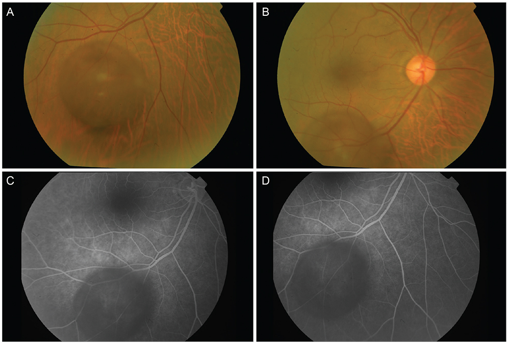
Fig. 1 There is a cyst floating inside the eye and partially masking the underlying the vasculature of the retina (A). There is a closer view of the cyst (B). In (C) and (D), there is fluorescein angiography (FA) of the fundus showing no vascularisation inside and on the surface of the cyst. FA also, shows a clear-edged hypofluorescence due to a pre-retinal masking effect.
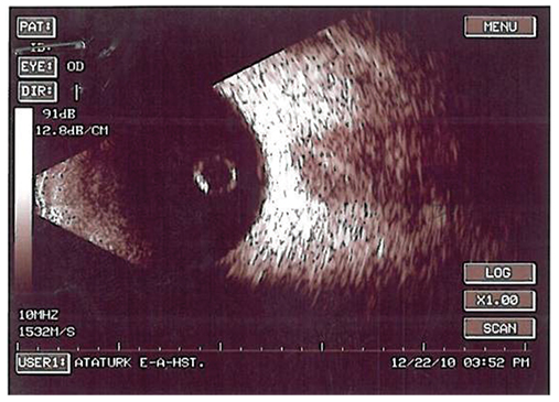
Fig. 2 B-mode ultrasonography of the cyst located in the posterior vitreous. There is no any attachments to the internal structures of the eye.
Reference
-
1. Cruciani F, Santino G, Salandri AG. Monolateral idiopathic cyst of the vitreous. Acta Ophthalmol Scand. 1999; 77:601–603.2. Duke-Elder S. Normal and abnormal development. Part 2. Congenital deformities. System of ophthalmology. London: Henry Kimpton;1964. Vol. 3:p. 763.3. Bayraktar Z, Kapran Z, Ozdogan S. Pigmented congenital vitreous cyst. Eur J Ophthalmol. 2004; 14:156–158.4. Awan KJ. Multiple free floating vitreous cysts with congenital nystagmus and esotropia. J Pediatr Ophthalmol. 1975; 12:49–53.5. Wolter JR, Martony CL, Smith C. A free-floating vitreous cyst in the otherwise normal eye of a young man. J Pediatr Ophthalmol. 1975; 12:243–245.6. Nork TM, Millecchia LL. Treatment and histopathology of a congenital vitreous cyst. Ophthalmology. 1998; 105:825–830.7. Jones WL. Free-floating vitreous cyst. Optom Vis Sci. 1998; 75:171–173.
