J Korean Ophthalmol Soc.
2014 Oct;55(10):1549-1553. 10.3341/jkos.2014.55.10.1549.
A Case of Isolated Orbital Sarcoidosis
- Affiliations
-
- 1Department of Ophthalmology, Daegu Fatima Hospital, Daegu, Korea. mskwak66@hotmail.com
- KMID: 2216877
- DOI: http://doi.org/10.3341/jkos.2014.55.10.1549
Abstract
- PURPOSE
The authors report a case of orbital sarcoidosis without evidence of systemic involvement.
CASE SUMMARY
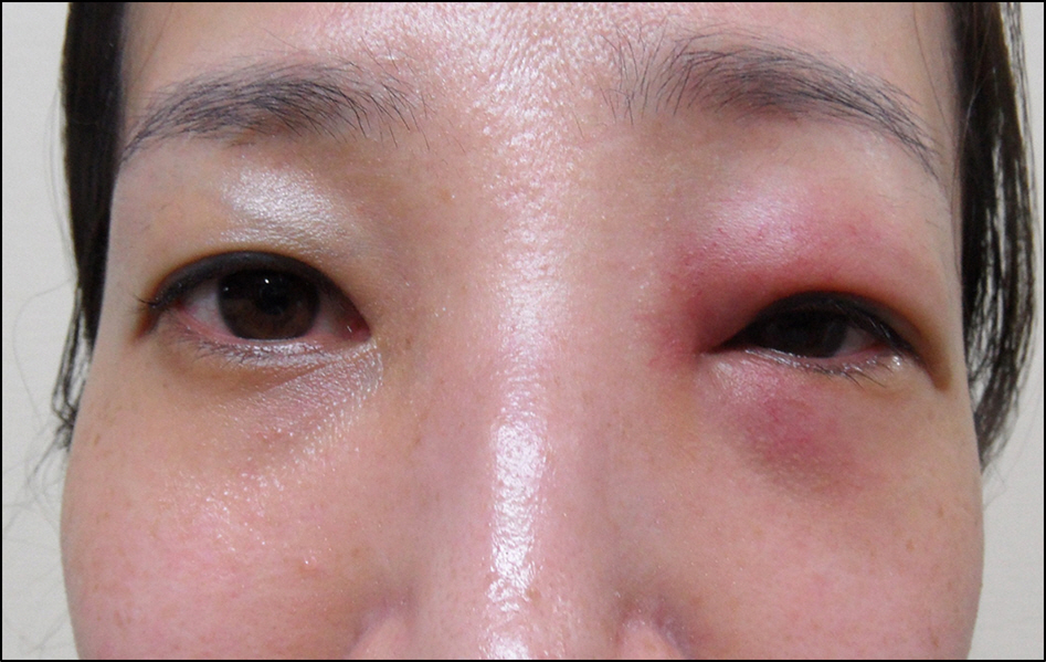
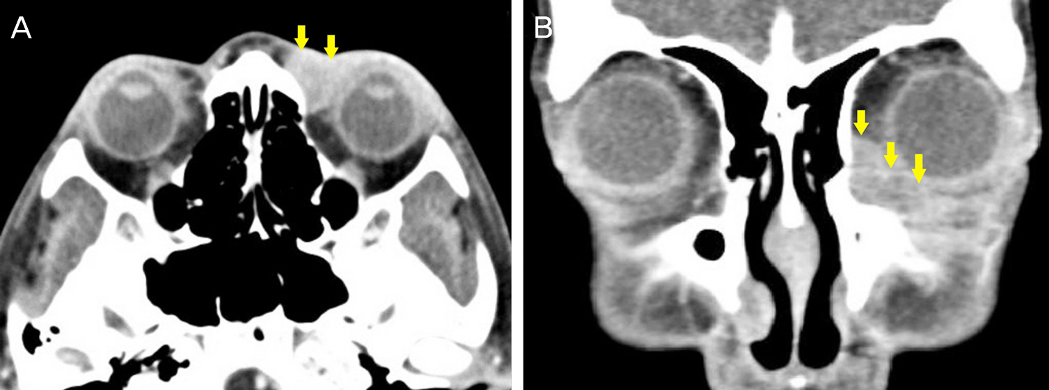
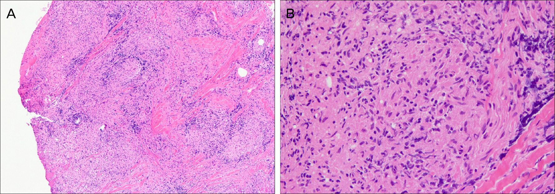
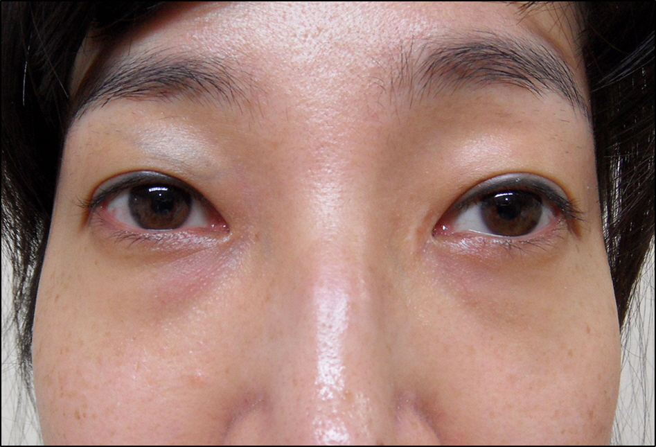
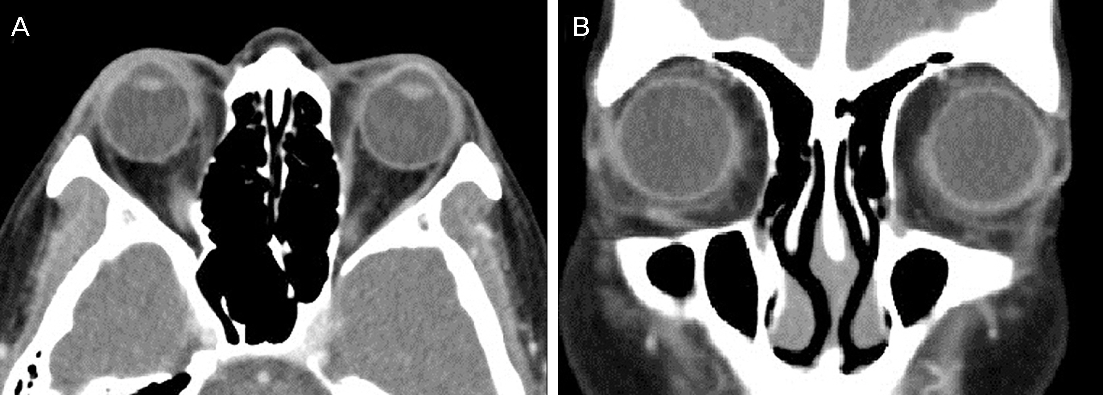
A 33-year-old female had a 1 month history of erythematous eyelid swelling. On physical examination, a firm and non-tender mass was observed diffusely along the upper, lower and medial canthal areas. A computed tomography (CT) scan showed a diffuse mass in the anterior orbit. We performed an incisional biopsy and histopathological examination revealed non-caseating granulomas and no evidence of a foreign body. Acid-fast-bacilli (AFB), methenamine silver and periodic-acid-schiff (PAS) stain showed no evidence of infection and chest radiograph was normal. Polymerase chain reaction (PCR) and interferon gamma secretion test showed no evidence of tuberculosis. Antinuclear antibody (ANA) and antineutrophil cytoplasmic antibody (ANCA) were negative and angiotensin converting enzyme (ACE) was within the normal range. Further systemic evaluations were compatible with a diagnosis of orbital sarcoidosis and oral prednisone was prescribed. Six weeks later, the erythematous eyelid swelling had disappeared and there was no evidence of recurrence to date.
MeSH Terms
-
Adult
Antibodies, Antineutrophil Cytoplasmic
Antibodies, Antinuclear
Biopsy
Diagnosis
Eyelids
Female
Foreign Bodies
Granuloma
Humans
Interferons
Methenamine
Orbit*
Peptidyl-Dipeptidase A
Physical Examination
Polymerase Chain Reaction
Prednisone
Radiography, Thoracic
Recurrence
Reference Values
Sarcoidosis*
Tuberculosis
Antibodies, Antineutrophil Cytoplasmic
Antibodies, Antinuclear
Interferons
Methenamine
Peptidyl-Dipeptidase A
Prednisone
Figure
Reference
-
References
1. MAYOCK RL, BERTRAND P, MORRISON CE, SCOTT JH. MANIFESTATIONS OF SARCOIDOSIS. ANALYSIS OF 145 PATIEN TS, WITH A REVIEW OF NINE SERIES SELECTED FROM THE LITERATURE. Am J Med. 1963; 35:67–89.2. Obenauf CD, Shaw HE, Sydnor CF, Klintworth GK. Sarcoidosis and its ophthalmic manifestations. Am J Ophthalmol. 1978; 86:648–55.
Article3. Demirci H, Christianson MD. Orbital and adnexal involvement in sarcoidosis: analysis of clinical features and systemic disease in 30 cases. Am J Ophthalmol. 2011; 151:1074–80.e1.
Article4. Liu CH, Ku WJ, Chang LC, et al. Idiopathic granulomatous orbital inflammation. Chang Gung Med J. 2003; 26:847–50.5. Lee DY, Kim DH, Yu SY, Kwak HW. A case of sarcoidosis misdiagnosed as tuberculosis in the early phase. J Korean Ophthalmol Soc. 2004; 45:438–43.6. Cho NC, Ahn HS. A case of palpebral sarcoidosis associated with granulomatous uveitis. J Korean Ophthalmol Soc. 1990; 31:819–23.7. Lee TH, Kim YJ, Sin DH. Case report: Ocular sareoidoasis. J Korean Ophthalmol Soc. 1993; 34:687–91.8. Kim KS, Choi BC, Kim HJ, et al. A case of sarcoidosis associated with uveitis and vitreous hemorrhage. J Korean Ophthalmol Soc. 1988; 29:433–42.9. Sohn MS, Kim CD, Cha OJ, et al. A case of sarcoidosis associated with granulomatous uveitis. J Korean Ophthalmol Soc. 1967; 8:11–6.10. Ko DA, Kim BJ. Sarcoidosis, presented as recurrent eyelid masses. J Korean Ophthalmol Soc. 2004; 45:1590–5.11. You IC, Moon HJ, Mun GH, et al. A case of sarcoidosis presented as multiple conjunctival and nasal mucosal nodule. J Korean Ophthalmol Soc. 2008; 49:1000–6.
Article12. Lee JK, Moon NJ. Orbital sarcoidosis presenting as diffuse swelling of the lower eyelid. Korean J Ophthalmol. 2013; 27:52–4.
Article13. Snead JW, Seidenstein L, Knific RJ, et al. Isolated orbital sarcoidosis as a cause for blepharoptosis. Am J Ophthalmol. 1991; 112:739–40.
Article14. Bäumer I, Zissel G, Schlaak M, Müller-Quernheim J. Th1/Th2 cell distribution in pulmonary sarcoidosis. Am J Respir Cell Mol Biol. 1997; 16:171–7.
Article15. Kim TW, Chung H, Yu HG. Clinical features in Korean patients with sarcoid uveitis. J Korean Ophthalmol Soc. 2008; 49:1483–90.
Article16. Hillerdal G, Nöu E, Osterman K, Schmekel B. Sarcoidosis: epidemiology and prognosis. A 15-year European study. Am Rev Respir Dis. 1984; 130:29–32.17. Weinreb RN, Tessler H. Laboratory diagnosis of ophthalmic sarcoidosis. Surv Ophthalmol. 1984; 28:653–64.
Article18. Jones NP. Sarcoidosis and uveitis. Ophthalmol Clin North Am. 2002; 15:319–26. vi.
Article19. Power WJ, Neves RA, Rodriguez A, et al. The value of combined serum angiotensin-converting enzyme and gallium scan in diagnosing ocular sarcoidosis. Ophthalmology. 1995; 102:2007–11.
Article20. Segal EI, Tang RA, Lee AG, et al. Orbital apex lesion as the presenting manifestation of sarcoidosis. J Neuroophthalmol. 2000; 20:156–8.
Article21. Bonfioli AA, Orefice F. Sarcoidosis. Semin Ophthalmol. 2005; 20:177–82.
Article22. Prabhakaran VC, Saeed P, Esmaeli B, et al. Orbital and adnexal sarcoidosis. Arch Ophthalmol. 2007; 125:1657–62.
Article23. Seamone CD, Nozik RA. Sarcoidosis and the eye. Ophthalmol Clin N Am. 1992; 5:567–76.

