A Case of Huge Pilocytic Astrocytoma Causing Eyeball Subluxation
- Affiliations
-
- 1Department of Ophthalmology and Visual Science, The Catholic University of Korea College of Medicine, Seoul, Korea. yswoph@hanmail.net
- KMID: 2216876
- DOI: http://doi.org/10.3341/jkos.2014.55.10.1543
Abstract
- PURPOSE
To report a relatively rare case of huge pilocytic astrocytoma of the optic nerve and optic chiasm causing eyeball subluxation.
CASE SUMMARY
An eight-year-old male presented with proptosis and visual loss in the left eye for one year. The radiological findings showed a 2.9 x 2.7 x 4.2-cm tumor on the left optic nerve and optic chiasm. For diagnosis and treatment, the patient underwent tumor resection and enucleation. Pathohistological analysis of the tumor specimen revealed pilocytic astrocytoma, which is classified by the World Health Organization as a grade I astrocytic tumor.
CONCLUSIONS
Astrocytoma is a tumor of the brain that affects children more often than adults. In general, gross-total resection of pilocytic astrocytoma is expected to be curative due to the non-invasive feature of the tumor. Considering pilocytic astrocytoma as differential diagnosis of orbital tumor in children with symptoms of rapidly progressive proptosis and decreased visual acuity is important because occurrence in the optic nerve and optic chiasm is possible.
Keyword
MeSH Terms
Figure
-
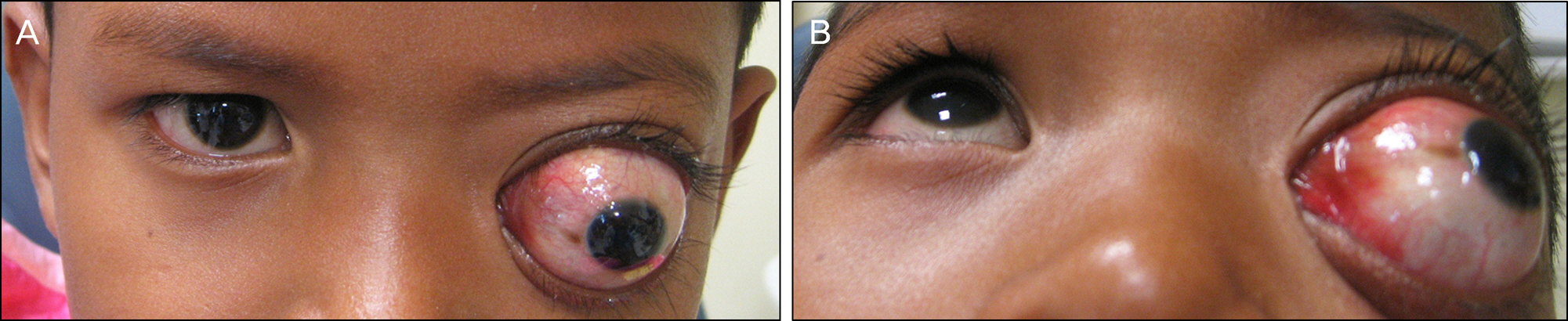
Figure 1. (A) Photograph of the patient taken at the first visit to the hospital shows severe proptosis and subluxation of the left eye. (B) Left eyeball is infero-laterally deviated (40-PD LXT, LHT by Hirschberg test).
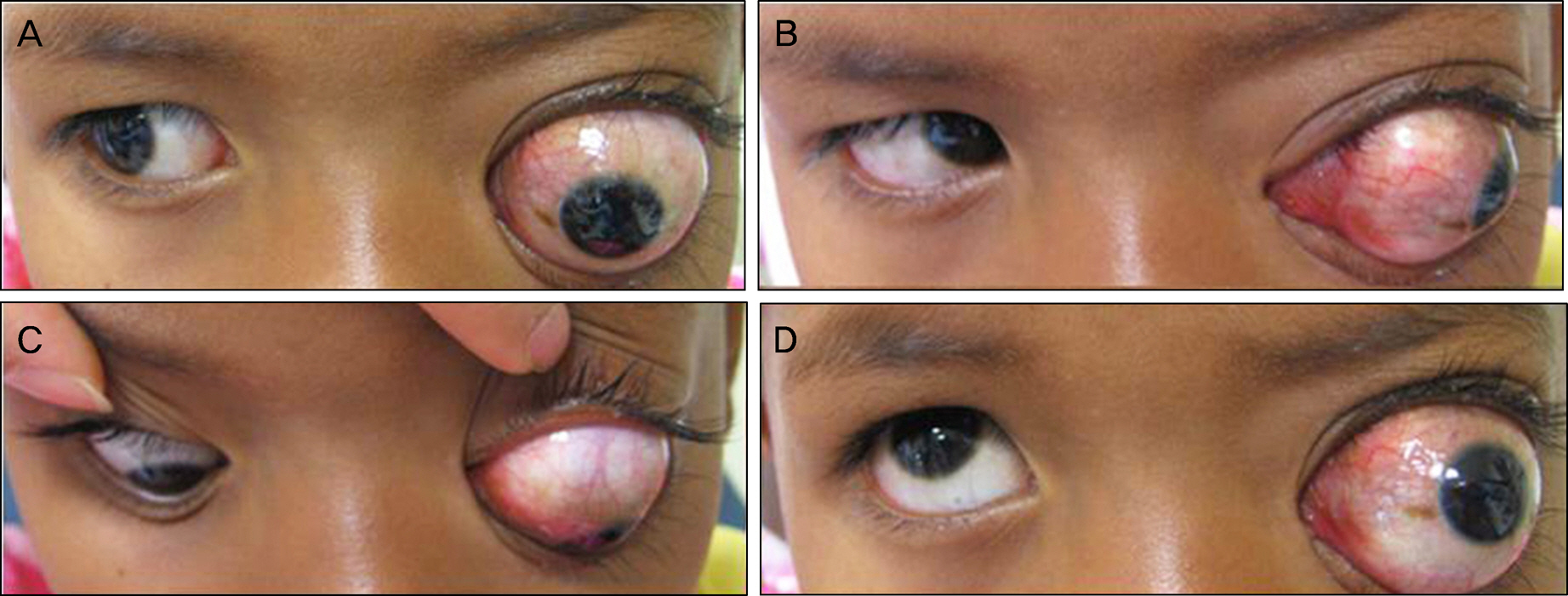
Figure 2. Photographs show limitation of the left eyeball movement for adduction and upward gaze. (A) Right side gaze, (B) left side gaze, (C) downward gaze, (D) upward gaze.
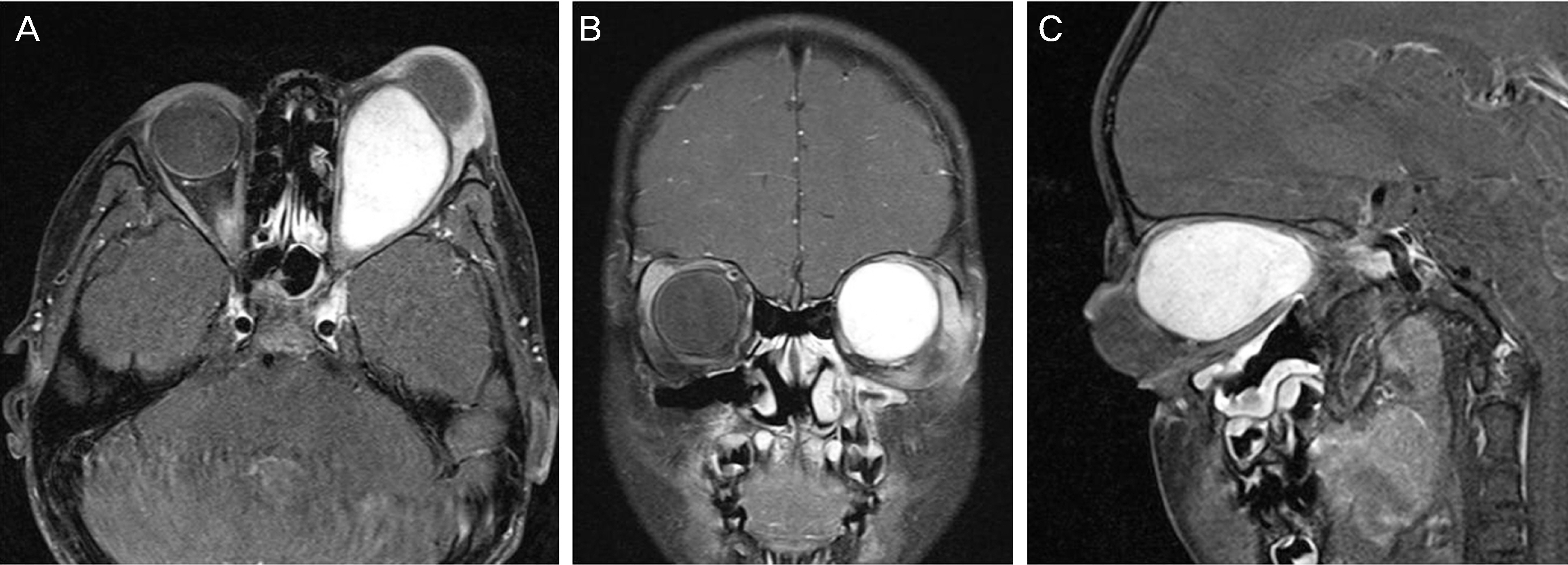
Figure 3. (A) 2.9 × 2.7 × 4.2-cm-sized enhancing mass involving the left optic nerve and optic chiasm (Orbit MRI (T1-weighted fat-suppressed gadolinium-enhanced image): A: axial view; B: coronal view; C: sagittal view).

Figure 4. Gross specimen of the optic nerve and optic chiasm astrocytoma, measuring 3.3 × 3.0 × 2.3 cm.
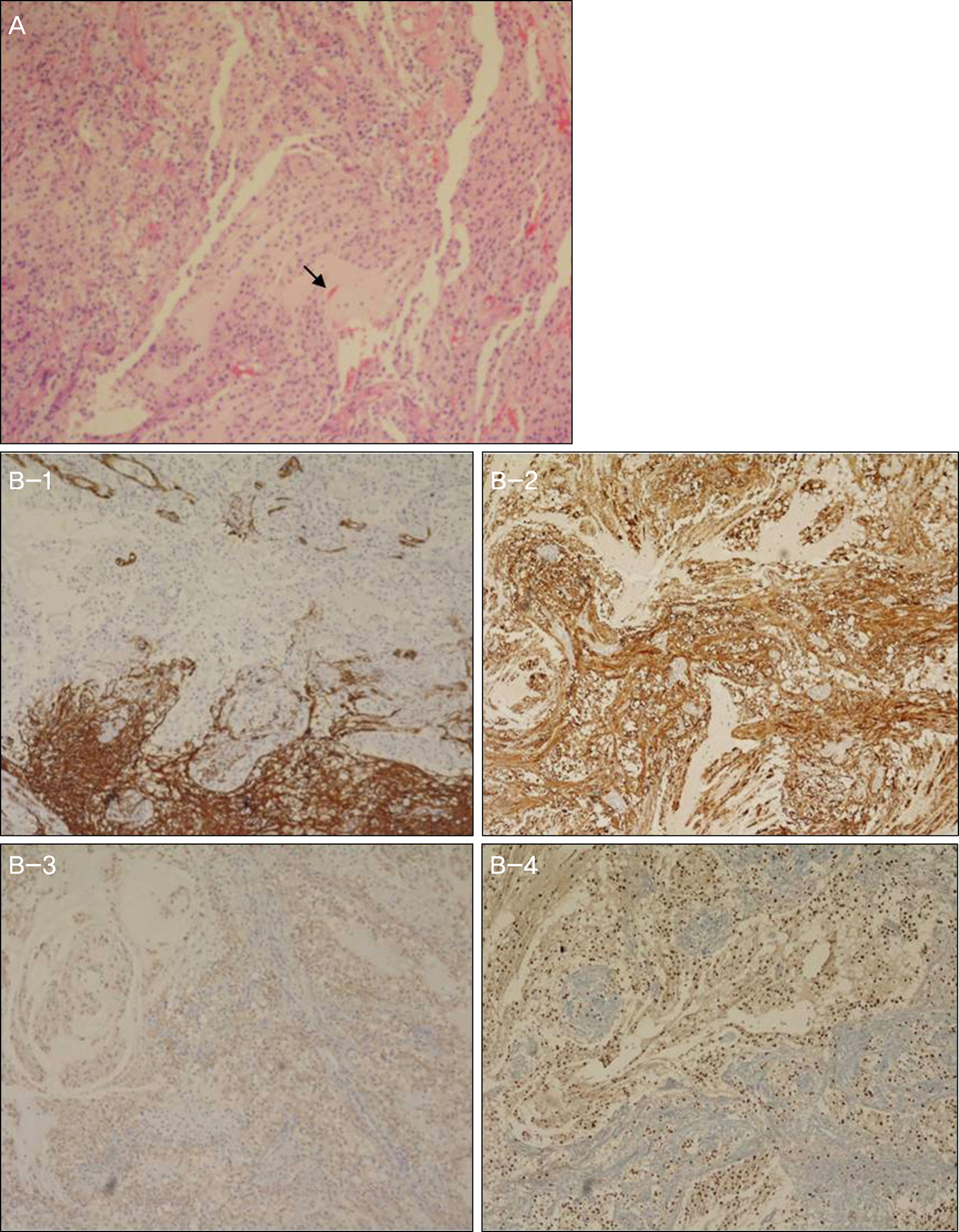
Figure 5. (A) Round and piloid cells constitute this tumor; Rosenthal fibers (arrow) is seen (H&E stain, ×100). (B) Immunohistochemical stains (×100), positive for (B-1) CD34, (B-2) Glial Fibrillary Acidic Protein (GFAP), (B-3) Synapto- physin, (B-4) Oligodendrocyte transcription factor (OLIG-2).
Reference
-
References
1. Louis DN, Ohgaki H, Wiestler OD, et al. The 2007 WHO classification of tumours of the central nervous system. Acta Neuropathol. 2007; 114:97–109.
Article2. Hong CS, Lehman NL, Sauvageau E. A pilocytic astrocytoma mimicking a clinoidal meningioma. Case Rep Radiol. 2014; 2014:524574.
Article3. Rodriguez FJ, Scheithauer BW, Burger PC, et al. Anaplasia in pilocytic astrocytoma predicts aggressive behavior. Am J Surg Pathol. 2010; 34:147–60.
Article4. Burkhard C, Di Patre PL, Schüler D, et al. A population-based study of the incidence and survival rates in patients with pilocytic astrocytoma. J Neurosurg. 2003; 98:1170–4.
Article5. Kim TJ, Cha SH, Chung YH. A case of a huge astrocytoma of the optic nerve. J Korean Ophthalmol Soc. 1983; 24:939–43.6. Chung YJ, Kim WK. A case of optic nerve astrocytoma. J Korean Ophthalmol Soc. 1985; 26:903–7.7. Jang JH, Ra YS, Kim JH, et al. The role of adjunctive therapy of optic pathway glioma in children. J Korean Neurosurg Soc. 2004; 35:136–41.8. Gillett GR, Symon L. Hypothalamic glioma. Surg Neurol. 1987; 28:291–300.
Article9. Silva MM, Goldman S, Keating G, et al. Optic pathway hypothalamic gliomas in children under three years of age: the role of chemotherapy. Pediatr Neurosurg. 2000; 33:151–8.
Article10. Binning MJ, Liu JK, Kestle JR, et al. Optic pathway gliomas: a review. Neurosurg Focus. 2007; 23:E2.
Article11. DODGE HW Jr, LOVE JG, CRAIG WM, et al. Gliomas of the optic nerves. AMA Arch Neurol Psychiatry. 1958; 79:607–21.
Article12. Rush JA, Younge BR, Campbell RJ, MacCarty CS. Optic glioma. Long-term follow-up of 85 histopathologically verified cases. Ophthalmology. 1982; 89:1213–9.13. Doh HJ, Kim IM, Lee CY, et al. Management of chiasmatic-hypothalamic gliomas in children. Journal of Korean Neurosurgical Society. 2001; 30:1115–9.14. Rosser T, Packer RJ. Intracranial neoplasms in children with neurofibromatosis 1. J Child Neurol. 2002; 17:630–7.15. Listernick R, Charrow J, Greenwald M, Mets M. Natural history of optic pathway tumors in children with neurofibromatosis type 1: a longitudinal study. J Pediatr. 1994; 125:63–6.
Article16. Grabenbauer GG, Schuchardt U, Buchfelder M, et al. Radiation therapy of optico-hypothalamic gliomas (OHG)–radiographic response, vision and late toxicity. Radiother Oncol. 2000; 54:239–45.17. Packer RJ, Sutton LN, Bilaniuk LT, et al. Treatment of chiasmatic/hypothalamic gliomas of childhood with chemotherapy: an update. Ann Neurol. 1988; 23:79–85.
Article18. Wisoff JH, Abbott R, Epstein F. Surgical management of exophytic chiasmatic-hypothalamic tumors of childhood. J Neurosurg. 1990; 73:661–7.
Article19. Hoffman HJ, Soloniuk DS, Humphreys RP, et al. Management and outcome of low-grade astrocytomas of the midline in children: a retrospective review. Neurosurgery. 1993; 33:964–71.20. Petronio J, Edwards MS, Prados M, et al. Management of chiasmal and hypothalamic gliomas of infancy and childhood with chemotherapy. J Neurosurg. 1991; 74:701–8.
Article21. Siffert J, Allen JC. Late effects of therapy of thalamic and hypothalamic tumors in childhood: vascular, neurobehavioral and neoplastic. Pediatr Neurosurg. 2000; 33:105–11.
Article22. Hufnagel TJ, Kim JH, Lesser R, et al. Malignant glioma of the optic chiasm eight years after radiotherapy for prolactinoma. Arch Ophthalmol. 1988; 106:1701–5.
Article23. Lee JW, Chung NG. The role of chemotherapy in the treatment of pediatric brain tumors. J Korean Med Assoc. 2012; 55:420–9.
Article
- Full Text Links
-
- Actions
-
Cited
- CITED
-
- Close
- Share
-
- Similar articles
-
- Multiple Solid Pilocytic Astrocytomas in Cerebellum with Neurofibromatosis Type I: A Case Report
- Supratentorial Pilocytic Astrocytoma Mimicking Convexity Meningioma with Early Anaplastic Transformation: A Case Report
- Pilocytic Astrocytoma Occuring in a Patient Treated with Gamma Knife Surgery: Case Report
- Juvenile Pilocytic Astrocytoma of the Hypohtalamus: Case Report
- Intracranial Hemorrhage from Pilocytic Astrocytoma in Pons
