Uncommon Ocular Manifestations of Neurofibromatosis: Case Report and Review
- Affiliations
-
- 1Department of Ophthalmology, Inha University School of Medicine, Incheon, Korea. ksm0724@medimail.co.kr
- KMID: 2215969
- DOI: http://doi.org/10.3341/jkos.2012.53.8.1200
Abstract
- PURPOSE
To report and review several cases of uncommon ocular manifestations in neurofibromatosis patients.
CASE SUMMARY
A 19-year-old woman diagnosed with type 2 neurofibromatosis visited our hospital with amblyopia of the right eye and mild visual disturbance of the left eye. Best corrected visual acuity was 20/250 in the right eye, 20/25 in the left eye and relative afferent pupillary defect in the right eye was observed. Fundus examination of both eyes showed papilledema. Magnetic resonance imaging showed schwannoma from the optic nerve to the optic chiasm. A 28-year-old woman diagnosed with type 2 neurofibromatosis visited our hospital with amblyopia of the right eye. Best corrected visual acuity was finger count in the right eye, 20/20 in the left eye and relative afferent pupillary defect in the right eye was observed. Fundus examination of the right eye showed a slightly elevated lesion at the macula, as well as dragged optic disc and retinal vessels to the macula. An 8-year-old girl diagnosed with type 1 neurofibromatosis visited our hospital with enophthalmos and strabismus of the left eye. On exophthalmometry, enophthalmos in the left eye was found; measurements were 15.0 mm in the right eye and 13.0 mm in the left eye. Three-dimensional computed tomography revealed sphenoidal hypoplasia and a left lateral orbital wall defect.
CONCLUSIONS
The authors of the present study report on neurofibromatosis patients who had an uncommon ocular manifestation. Neurofibromatosis can represent various ocular manifestations but reports of compressive optic neuropathy, dragged disc syndrome and sphenoidal hypoplasia are rare.
Keyword
MeSH Terms
Figure
-

Figure 1 Case 1. The fundus photographs of 19-year-old female with type 2 neurofibromatosis. Fundus examination shows papilledema in both eyes and peripapillary retinal hemorrhage in the right eye.
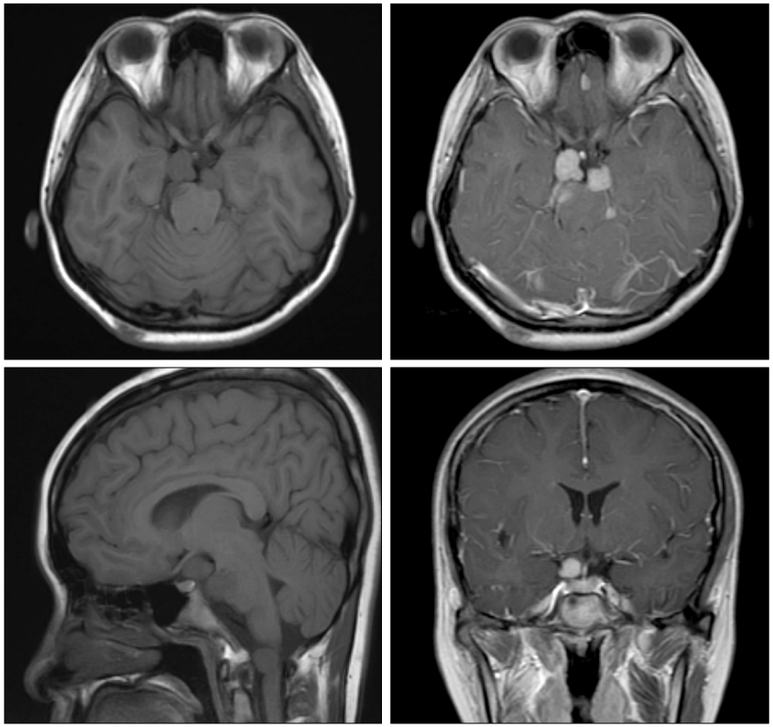
Figure 2 Case 1. Magnetic resonance imaging. Two Schwannomas were noted nearby optic nerve to optic chiasm, each measured about 27.0 × 12.0 mm, 23.0 × 10.0 mm in axial T1 image and axial T1 Gd enhance image. In T1 sagittal view, round mass is noted just inferior to the right optic nerve. In the T1 coronal Gd-enhanced image, round mass is also noted just inferior to the optic chiasm.
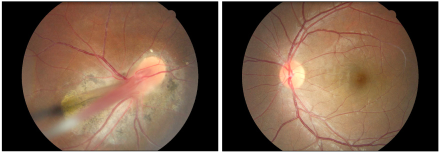
Figure 3 Case 2. The fundus photographs of 28-year-old female with type 2 neurofibromatosis. Fundus examination of the right eye shows abnormal temporal dragging of optic disc and retinal vessels and slightly elevated lesion at the macula and geographic chorioretinal atrophy at the posterior pole. Fundus examination of the left eye shows a normal fundus.
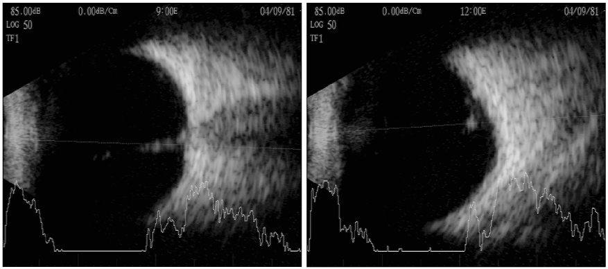
Figure 4 Case 2. The ultrasonography of the right eye shows a 4 mm-sized protruding mass into the vitreous cavity with high echogenicity in the lateral and superior views.

Figure 5 Case 3. The photographs of nine cardinal directions of gaze of 8-year-old female with type 2 neurofibromatosis. At the primary gaze, the enophthalmos and pseudoesotropia of the left eye is noted.
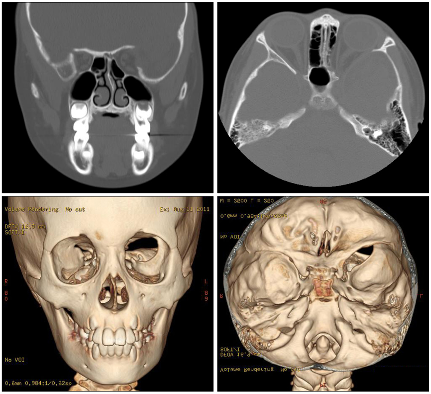
Figure 6 Case 3. Multi-Detector Computed Tomography. Facial CT scan illustrates the sphenoidal hypoplasia, left lateral orbital wall defect and enlarged middle cranial fossa. Three dimensional reconstructions of computed tomography images illustrate the enlargement of the left superior orbital fissure.
Reference
-
1. Suh YW, Oh CH, Kim HM. Subconjunctival juvenile xanthogranuloma involving the limbus associated with neurofibromatosis. J Korean Ophthalmol Soc. 2005. 46:1733–1736.2. Bang JS, Yang HS, Ahn JH, et al. Ophthalmic manifestations in patients with neurofibromatosis. J Korean Ophthalmol Soc. 2008. 49:1829–1838.3. Duke-Elder S. System of Ophthalmology. 1967. vol. 10:15th ed. London: Henry Kimpton;738.4. Riccardi VM. Neurofibromatosis: clinical heterogeneity. Curr Probl Cancer. 1982. 7:1–34.5. Lee KH, Park JW, Yoon KC. A case of neurofibromatosis of the orbit and ocular surface. J Korean Ophthalmol Soc. 2007. 48:1276–1280.6. Lammert M, Friedman JM, Kluwe L, Mautner VF. Prevalence of neurofibromatosis 1 in German children at elementary school enrollment. Arch Dermatol. 2005. 141:71–74.7. Evans DG, Moran A, King A, et al. Incidence of vestibular schwannoma and neurofibromatosis 2 in the North West of England over a 10-year period: higher incidence than previously thought. Otol Neurotol. 2005. 26:93–97.8. Evans DG, Howard E, Giblin C, et al. Birth incidence and prevalence of tumor-prone syndromes: estimates from a UK family genetic register service. Am J Med Genet A. 2010. 152A:327–332.9. Antinheimo J, Sankila R, Carpén O, et al. Population-based analysis of sporadic and type 2 neurofibromatosis-associated meningiomas and schwannomas. Neurology. 2000. 54:71–76.10. Ledbetter DH, Rich DC, O'Connell P, et al. Precise localization of NF1 to 17q11.2 by balanced translocation. Am J Hum Genet. 1989. 44:20–24.11. Feldkamp MM, Gutmann DH, Guha A. Neurofibromatosis type 1: piecing the puzzle together. Can J Neurol Sci. 1998. 25:181–191.12. Seizinger BR, Martuza RL, Gusella JF. Loss of genes on chromosome 22 in tumorigenesis of human acoustic neuroma. Nature. 1986. 322:644–647.13. Savar A, Cestari DM. Neurofibromatosis type I: genetics and clinical manifestations. Semin Ophthalmol. 2008. 23:45–51.14. Bosch MM, Boltshauser E, Harpes P, Landau K. Ophthalmologic findings and long-term course in patients with neurofibromatosis type 2. Am J Ophthalmol. 2006. 141:1068–1077.15. Gaonker CH, Mukherjee AK, Pokle M. Involvement of the eye and orbit in neurofibromatosis type 1. Indian J Ophthalmol. 1992. 40:2–4.16. Huson S, Jones D, Beck L. Ophthalmic manifestations of neurofibromatosis. Br J Ophthalmol. 1987. 71:235–238.17. Good WV, Erodsky MC, Edwards MS, Hoyt WF. Bilateral retinal hamartomas in neurofibromatosis type 2. Br J Ophthalmol. 1991. 75:190.18. Hwang JM, Jung YC. The etiology of optic neuropathy. J Korean Ophthalmol Soc. 1999. 40:1078–1083.19. Laemmer R, Heckmann JG, Mardin CY, et al. Detection of nerve fiber atrophy in apparently effectively treated papilledema in idiopathic intracranial hypertension. Graefes Arch Clin Exp Ophthalmol. 2010. 248:1787–1793.20. Lewis RA, Gerson LP, Axelson KA, et al. von Recklinghausen neurofibromatosis. II. Incidence of optic gliomata. Ophthalmology. 1984. 91:929–935.21. Listernick R, Charrow J, Gutmann DH. Intracranial gliomas in neurofibromatosis type 1. Am J Med Genet. 1999. 89:38–44.22. Guillamo JS, Créange A, Kalifa C, et al. Prognostic factors of CNS tumours in Neurofibromatosis 1 (NF1): a retrospective study of 104 patients. Brain. 2003. 126(Pt 1):152–160.23. Créange A, Zeller J, Rostaing-Rigattieri S, et al. Neurological complications of neurofibromatosis type 1 in adulthood. Brain. 1999. 122(Pt 3):473–481.24. Parry DM, Eldridge R, Kaiser-Kupfer MI, et al. Neurofibromatosis 2 (NF2): clinical characteristics of 63 affected individuals and clinical evidence for heterogeneity. Am J Med Genet. 1994. 52:450–461.25. Mautner VF, Lindenau M, Baser ME, et al. The neuroimaging and clinical spectrum of neurofibromatosis 2. Neurosurgery. 1996. 38:880–885.26. Evans DG, Huson SM, Donnai D, et al. A clinical study of type 2 neurofibromatosis. Q J Med. 1992. 84:603–618.27. Schick U, Bleyen J, Hassler W. Treatment of orbital schwannomas and neurofibromas. Br J Neurosurg. 2003. 17:541–545.28. Chua CN, Alhady M, Ngo CT, et al. Solitary nasal neurofibroma presenting as compressive optic neuropathy. Eye (Lond). 2006. 20:1406–1408.29. Civit T, Freppel S. [Intraorbital schwannomas and solitary neurofibromas]. Neurochirurgie. 2010. 56:137–141.30. Evans DG. Neurofibromatosis type 2 (NF2): a clinical and molecular review. Orphanet J Rare Dis. 2009. 4:16.31. Evans DG. Neurofibromatosis 2 [Bilateral acoustic neurofibromatosis, central neurofibromatosis, NF2, neurofibromatosis type II]. Genet Med. 2009. 11:599–610.32. Ragge NK, Baser ME, Klein J, et al. Ocular abnormalities in neurofibromatosis 2. Am J Ophthalmol. 1995. 120:634–641.33. McLaughlin ME, Pepin SM, Maccollin M, et al. Ocular pathologic findings of neurofibromatosis type 2. Arch Ophthalmol. 2007. 125:389–394.34. Ragge NK, Baser ME, Riccardi VM, Falk RE. The ocular presentation of neurofibromatosis 2. Eye (Lond). 1997. 11(Pt 1):12–18.35. Kroll P, Wiegand W, Schmidt J. Vitreopapillary traction in proliferative diabetic vitreoretinopathy. Br J Ophthalmol. 1999. 83:261–264.36. Karatas M, Ramirez JA, Ophir A. Diabetic vitreopapillary traction and macular oedema. Eye (Lond). 2005. 19:676–682.37. Rumelt S, Karatas M, Pikkel J, et al. Optic disc traction syndrome associated with central retinal vein occlusion. Arch Ophthalmol. 2003. 121:1093–1097.38. Grant EA, Trzupek KM, Reiss J, et al. Combined retinal hamartomas leading to the diagnosis of neurofibromatosis type 2. Ophthalmic Genet. 2008. 29:133–138.39. Gicquel JJ, Vabres P, Mercié M, et al. [Dragged disc syndrome in a patient presenting neurofibromatosis type II: a case study]. J Fr Ophtalmol. 2005. 28:527–529.40. Vianna RN, Pacheco DF, Vasconcelos MM, de Laey JJ. Combined hamartoma of the retina and retinal pigment epithelium associated with neurofibromatosis type-1. Int Ophthalmol. 2001. 24:63–66.41. Landau K, Yargil GM. Ocular fundus in neurofibromatosis type 2. Br J Ophthalmol. 1993. 77:646–649.42. Pinna A, Demontis S, Maltese G, et al. Absence of the greater sphenoid wing in neurofibromatosis 1. Arch Ophthalmol. 2005. 123:1454.43. Onbas O, Aliagaoglu C, Calikoglu C, et al. Absence of a sphenoid wing in neurofibromatosis type 1 disease: imaging with multidetector computed tomography. Korean J Radiol. 2006. 7:70–72.44. Erb MH, Uzcategui N, See RF, Burnstine MA. Orbitotemporal neurofibromatosis: classification and treatment. Orbit. 2007. 26:223–228.45. Jo YJ, Lee SB, Kwon HJ, et al. Inducible dynamic proptosis in a neurofibromatosis patient with arachnoid cyst. J Korean Ophthalmol Soc. 2011. 52:93–96.
