ON and OFF Responses of the Electroretinogram in Patients with Glaucoma
- Affiliations
-
- 1Department of Ophthalmology, Soonchunhyang University College of Medicine, Bucheon, Korea. yhohn@schmc.ac.kr
- KMID: 2215952
- DOI: http://doi.org/10.3341/jkos.2012.53.8.1104
Abstract
- PURPOSE
To investigate whether there is a difference in ON- and OFF-responses of the photopic electroretinogram (ERG) in glaucomatous eyes.
METHODS
Photopic ERG and optical coherence tomography were performed in 15 normal, 13 glaucoma suspect, and 22 glaucoma subjects. Amplitudes and implicit times for a, b, d, i, photopic negative response (PhNR), and retinal nerve fiber layer thickness were compared among the three groups.
RESULTS
The PhNROFF amplitude (microV) was 19.05 +/- 11.41 in the glaucoma group, 14.24 +/- 10.37 in the glaucoma suspect group, and 2.69 +/- 12.16 in the normal group, demonstrating a significant difference among the three groups (p < 0.01). The PhNRON amplitude (microV) was 20.15 +/- 13.99 in the glaucoma group, 31.49 +/- 17.09 in the glaucoma suspect group, and 37.59 +/- 9.53 in the normal group, a significant difference (p < 0.01). However, there was no significant difference between the three groups. The ON-OFF response PhNR amplitude was correlated with retinal nerve fiber thickness (r = 0.481, r = -0.480, respectively), and areas under the receiver operating characteristic curve were 0.782, and 0.718, respectively.
CONCLUSIONS
There is a potential role for the ON-OFF response PhNR in early detection of glaucomatous damage.
MeSH Terms
Figure
-
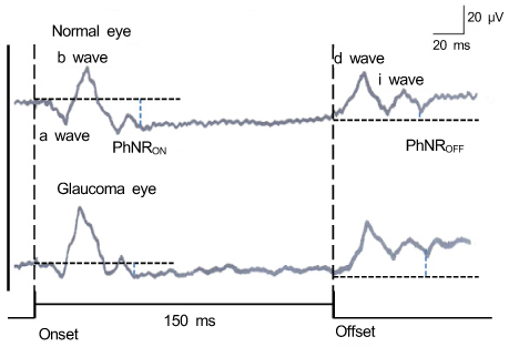
Figure 1 ON-OFF response electroretinogram show group-averaged responses from 15 normal subjects and 22 glaucoma patients. Photopic negative response of ON-response (PhNRON) is measured from the onset-level. Photopic negative response of OFF-response (PhNROFF) is measured from the offset level. Glaucoma patients show different amplitudes for the PhNRON, the PhNROFF in comparison with the normal eye.
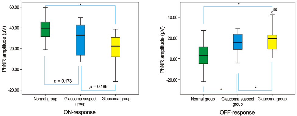
Figure 2 Comparison of Photopic negative response (PhNR) amplitude in the ON-response and OFF-response among the three groups. *p < 0.05 by ANOVA and Scheffe test was considered to be significant.

Figure 3 Correlation of the photopic negative response (PhNR) amplitude in the ON-OFF response and the retinal nerve fiber layer thickness among the three groups. *p < 0.05 by Pearson correlation test was considered to be significant. (A) PhNRON response. (B) PhNROFF response.

Figure 4 The receiver operating characteristic (ROC) curve of photopic negative response (PhNR) amplitude of ON-response and PhNR amplitude of OFF-response for discrimination of glaucoma from normal eyes.
Reference
-
1. Marmor MF, Fulton AB, Holder GE, et al. ISCEV standard for full-field clinical electroretinography (2008 update). Doc Ophthalmol. 2009. 118:69–77.2. Sieving PA. Photopic ON- and OFF-pathway abnormalities in retinal dystrophies. Trans Am Ophthalmol Soc. 1993. 91:701–773.3. Bush RA, Sieving PA. A proximal retinal component in the primate photopic ERG a-wave. Invest Ophthalmol Vis Sci. 1994. 35:635–645.4. Hood DC, Birch DG. Phototransduction in human cones measured using the alpha-wave of the ERG. Vision Res. 1995. 35:2801–2810.5. Sieving PA, Murayama K, Naarendorp F. Push-pull model of the primate photopic electroretinogram: a role for hyperpolarizing neurons in shaping the b-wave. Vis Neurosci. 1994. 11:519–532.6. Evers HU, Gouras P. Three cone mechanisms in the primate electroretinogram: two with, one without off-center bipolar responses. Vision Res. 1986. 26:245–254.7. Viswanathan S, Frishman LJ, Robson JG, et al. The photopic negative response of the macaque electroretinogram: reduction by experimental glaucoma. Invest Ophthalmol Vis Sci. 1999. 40:1124–1136.8. Drasdo N, Aldebasi YH, Chiti Z, et al. The s-cone PHNR and pattern ERG in primary open angle glaucoma. Invest Ophthalmol Vis Sci. 2001. 42:1266–1272.9. Colotto A, Falsini B, Salgarello T, et al. Photopic negative response of the human ERG: losses associated with glaucomatous damage. Invest Ophthalmol Vis Sci. 2000. 41:2205–2211.10. Viswanathan S, Frishman LJ, Robson JG, Walters JW. The photopic negative response of the flash electroretinogram in primary open angle glaucoma. Invest Ophthalmol Vis Sci. 2001. 42:514–522.11. Gotoh Y, Machida S, Tazawa Y. Selective loss of the photopic negative response in patients with optic nerve atrophy. Arch Ophthalmol. 2004. 122:341–346.12. Iester M, De Ferrari R, Zanini M. Topographic analysis to discriminate glaucomatous from normal optic nerve heads with a confocal scanning laser: new optic disk analysis without any observer input. Surv Ophthalmol. 1999. 44:Suppl 1. S33–S40.13. Gupta N, Weinreb RN. New definitions of glaucoma. Curr Opin Ophthalmol. 1997. 8:38–41.14. Weber AJ, Harman CD. Structure-function relations of parasol cells in the normal and glaucomatous primate retina. Invest Ophthalmol Vis Sci. 2005. 46:3197–3207.15. North RV, Jones AL, Drasdo N, et al. Electrophysiological evidence of early functional damage in glaucoma and ocular hypertension. Invest Ophthalmol Vis Sci. 2010. 51:1216–1222.16. Harwerth RS, Carter-Dawson L, Shen F, et al. Ganglion cell losses underlying visual field defects from experimental glaucoma. Invest Ophthalmol Vis Sci. 1999. 40:2242–2250.17. Sommer A, Katz J, Quigley HA, et al. Clinically detectable nerve fiber atrophy precedes the onset of glaucomatous field loss. Arch Ophthalmol. 1991. 109:77–83.18. Kondo M, Piao CH, Tanikawa A, et al. Amplitude decrease of photopic ERG b-wave at higher stimulus intensities in humans. Jpn J Ophthalmol. 2000. 44:20–28.19. Park SE, Chang JH, Choi KS, et al. Photopic ON- and OFF- responses in Korean normal subjects. J Korean Ophthalmol Soc. 2008. 49:471–478.20. Sustar M, Hawlina M, Brecelj J. ON- and OFF-response of the photopic electroretinogram in relation to stimulus characteristics. Doc Ophthalmol. 2006. 113:43–52.21. Hanitzsch R. ERG-Veränderungen an isolierten Netzhäuten glaukomerkrankter Augen. Graefe's Arch Clin Exp Ophthalmol. 1966. 170:342–348.22. Kondo M, Kurimoto Y, Sakai T, et al. Recording focal macular photopic negative response (PhNR) from monkeys. Invest Ophthalmol Vis Sci. 2008. 49:3544–3550.23. Horn FK, Gottschalk K, Mardin CY, et al. On and off responses of the photopic fullfield ERG in normal subjects and glaucoma patients. Doc Ophthalmol. 2011. 122:53–62.24. Ueno S, Kondo M, Ueno M, et al. Contribution of retinal neurons to d-wave of primate photopic electroretinograms. Vision Res. 2006. 46:658–664.25. Rangaswamy NV, Frishman LJ, Dorotheo EU, et al. Photopic ERGs in patients with optic neuropathies: comparison with primate ERGs after pharmacologic blockade of inner retina. Invest Ophthalmol Vis Sci. 2004. 45:3827–3837.26. Rosolen SG, Rigaudière F, LeGargasson JF, et al. Comparing the photopic ERG i-wave in different species. Vet Ophthalmol. 2004. 7:189–192.
- Full Text Links
-
- Actions
-
Cited
- CITED
-
- Close
- Share
-
- Similar articles
-
- Prognostic Factors for Neovascular Glaucoma after Vitrectomy in Eyes with Proliferative Diabetic Retinopathy
- Effect of Intracameral Mitomycin-C Irrigation on ERG in Rabbit
- The Clinical Applications of Multifocal Electroretinogram in Diabetic Retinopathy
- A Clinical Evaluation of Trabeculectomy
- A Correlation with Retinal Thickness Using Retinal Thickness Analyzer and the Responses of Multifocal Electroretinogram in Patients with Diabetic Macular Edema
