Treatment of Epiphora in Patients with Conjunctivochalasis Using Conjunctival Fixation to the Sclera
- Affiliations
-
- 1Department of Ophthalmology, Incheon St. Mary's Hospital, College of Medicine, The Catholic University of Korea, Incheon, Korea. Ny55@freechal.com
- KMID: 2215946
- DOI: http://doi.org/10.3341/jkos.2012.53.8.1063
Abstract
- PURPOSE
To report the effects and complications of conjunctival fixation to the sclera in conjunctivochalasis patients with inferior punctal occlusion.
METHODS
The authors of the present study evaluated the degree of conjunctivochalasis and performed Fluorescein Dye Disappearance Test (FDT) in 15 eyes of 8 patients diagnosed with conjunctivochalasis with inferior punctal occlusion. Under topical anesthesia, the inferior bulbar conjunctiva was attached to the sclera with 3 8-0 vicryl stitches 8 mm posterior from the limbus. After surgery, the relief of symptoms, postoperative complications and improvement of conjunctivochalasis were observed.
RESULTS
One week after the surgery, all 15 eyes achieved a subjective improvement of symptoms and the degree of conjunctivochalasis and FDT showed statistical difference after surgery (p = 0.000, 0.000, respectively). A complication occurred in 1 eye which was a retinal hemorrhage due to scleral puncture.
CONCLUSIONS
Conjunctival fixation to the sclera could improve epiphora in conjunctivochalasis patients with inferior punctal occlusion. However, this procedure should be performed with caution.
MeSH Terms
Figure
-

Figure 1 (A) Intraoperative photograph of the surgical procedure. To expose the inferior fornix, we placed a corneal traction suture with 8-0 Vicryl. (B) Postoperative photograph. Central suture material was observed about 8mm from the limbus (arrow).
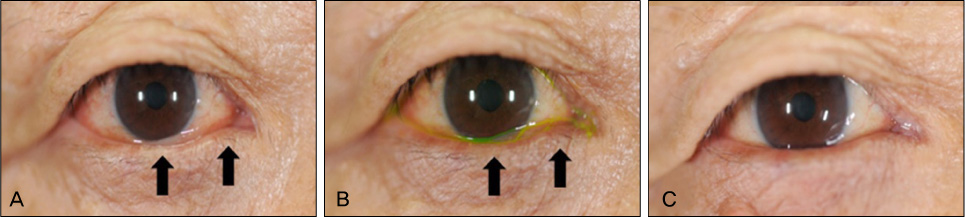
Figure 2 (A) A 74-year-old man with conjunctivochalasis of grade 2. Conjunctival folds on the central and medial lower eyelid margin were observed. (B) Redundant conjunctiva was seen definitely with fluorescein dye. (C) Seven months after surgery, no conjunctival folds were evident on the lower eyelid margin.
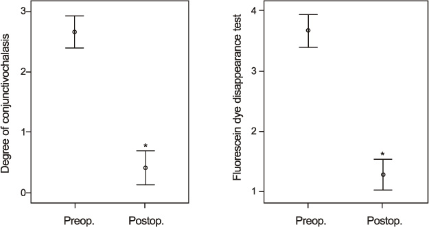
Figure 3 Graphs demonstrating the degree of conjunctivochalasis and fluorescein dye disappearance test preoperatively and postoperatively. Each dot represents the average of each factor (95% confidence interval). *Statistical significance (p < 0.05) compared with preoperative status.
Cited by 1 articles
-
Combination Surgery of Silicone Tube Intubation and Conjunctival Resection in Patients with Epiphora
Seon Tae Kim, Long Yu Jin, Hee Bae Ahn
Korean J Ophthalmol. 2018;32(6):438-444. doi: 10.3341/kjo.2018.0044.
Reference
-
1. Hughes WL. Conjunctivochalasis. Am J Ophthalmol. 1942. 25:48–51.2. Meller D, Tseng SC. Conjunctivochalasis: literature review and possible pathophysiology. Surv Ophthalmol. 1998. 43:225–232.3. Liu D. Conjunctivochalasis. A cause of tearing and its management. Ophthal Plast Reconstr Surg. 1986. 2:25–28.4. Byon D, Song CH, Shim JK, Shyn KH. Conjunctivochalasis as a cause of epiphora and its histopathological findings. J Korean Ophthalmol Soc. 1996. 37:400–404.5. Ko SM, Kim MK, Kim JC. The role of mast cell in hyperlaxity of conjunctiva. J Korean Ophthalmol Soc. 1997. 38:949–955.6. Otaka I, Kyu N. A new surgical technique for management of conjunctivochalasis. Am J Ophthalmol. 2000. 129:385–387.7. Lim HJ, Lee JK, Park DJ. Conjunctivochalasis surgery: Amniotic membrane transplantation with fibrin glue. J Korean Ophthalmol Soc. 2008. 49:195–204.8. Brodbaker E, Bahar I, Slomovic AR. Novel use of fibrin glue in the treatment of conjunctivochalasis. Cornea. 2008. 27:950–952.9. Park CK, Cho NC. A modified surgical technique for 2 cases of conjunctivochalasis near the lower punctum. J Korean Ophthalmol Soc. 1993. 34:75–78.10. Nam K, Jo YJ, Lee SB. The efficacy of fibrin glue in surgical treatment of conjunctivochalasis with epiphora. J Korean Ophthalmol Soc. 2010. 51:498–503.11. Kim HH, Shin DS, Lee KW. Effects of cauterization with suturing in treatment of conjunctivochalasis: 4 Cases. J Korean Ophthalmol Soc. 2006. 47:843–846.12. Meller D, Maskin SL, Pires RT, Tseng SC. Amniotic membrane transplantation for symptomatic conjunctivochalasis refractory to medical treatments. Cornea. 2000. 19:796–803.13. Firestone DE, Lauder AJ. Chemistry and mechanics of commonly used sutures and needles. J Hand Surg Am. 2010. 35:486–488.14. Pillai CK, Sharma CP. Review paper: absorbable polymeric surgical sutures: chemistry, production, properties, biodegradability, and performance. J Biomater Appl. 2010. 25:291–366.15. Höh H, Schirra F, Kienecker C, Ruprecht KW. [Lid-parallel conjunctival folds are a sure diagnostic sign of dry eye]. Ophthalmologe. 1995. 92:802–808.
- Full Text Links
-
- Actions
-
Cited
- CITED
-
- Close
- Share
-
- Similar articles
-
- The Efficacy of Fibrin Glue in Surgical Treatment of Conjunctivochalasis With Epiphora
- Treatment of Conjunctivochalasis Using Bipolar Cautery
- Effects of Cauterization with Suturing in Treatment of Conjunctivochalasis: 4 Cases
- The Effects of Conjunctival Shield on Pain Alleviation During Cataract Surgery in Conjunctivochalasis Patients
- A Modified Surgical Technique for 2 Cases of Conjunctivochalasis Near the Lower Punctum
