J Korean Ophthalmol Soc.
2010 Apr;51(4):498-503. 10.3341/jkos.2010.51.4.498.
The Efficacy of Fibrin Glue in Surgical Treatment of Conjunctivochalasis With Epiphora
- Affiliations
-
- 1Department of Ophthalmology, Chungnam National University College of medicine, Daejeon, Korea. sblee@cnu.ac.kr
- 2Research Institute for Medical Science, Chungnam National University, Daejeon, Korea.
- KMID: 2213389
- DOI: http://doi.org/10.3341/jkos.2010.51.4.498
Abstract
- PURPOSE
To investigate the efficacy of fibrin glue used in conjunctival resection for conjunctivochalasis with epiphora.
METHODS
Twenty-three patients (42 eyes) with conjunctivochalasis without nasolacrimal duct obstruction underwent conjunctival resection using either absorbable sutures (11 patients, 20 eyes, Group 1) or fibrin glue (12 patients, 22 eyes, Group 2) to attach the conjunctiva to the sclera. Outcomes recorded were improvement of epiphora, postoperative discomfort, and operation time. Postoperative discomfort was analyzed only in one eye (right eye) in case that the both eyes were operated.
RESULTS
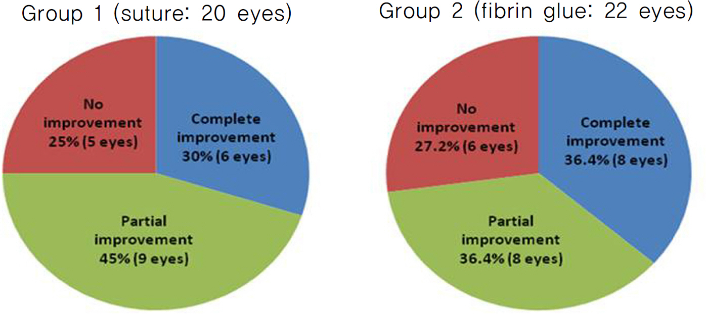
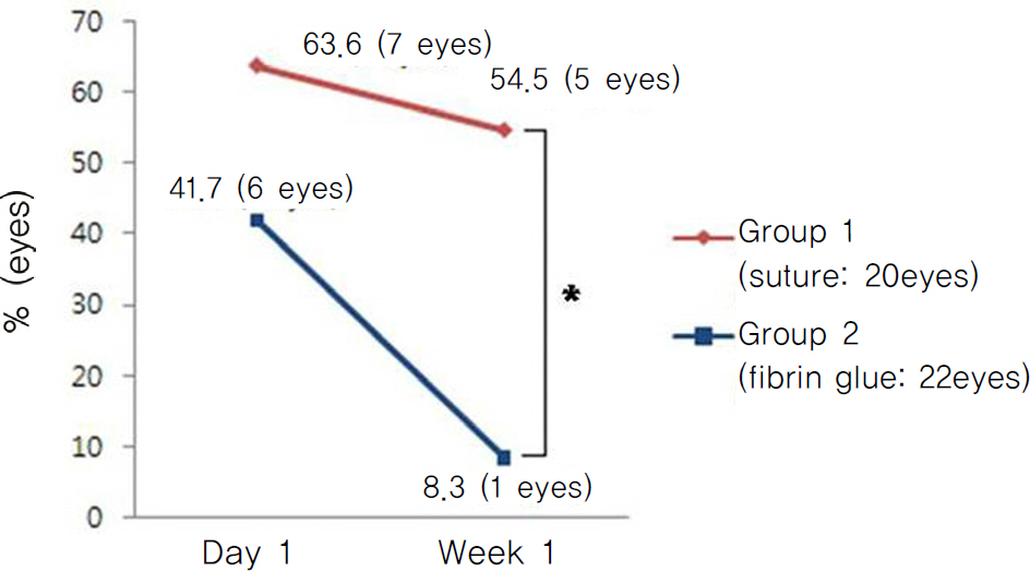
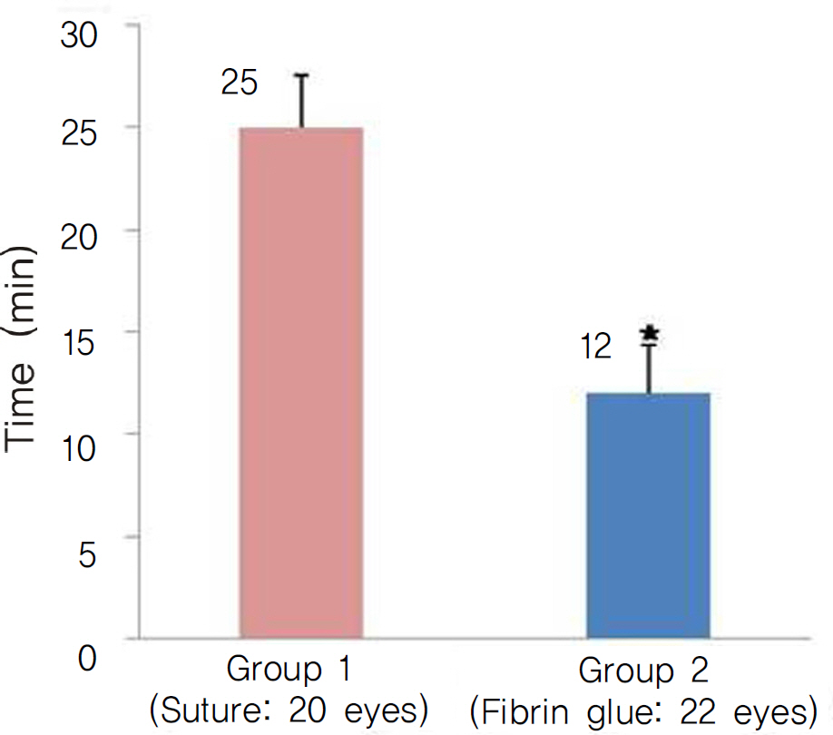
Epiphora completely improved in 6 eyes (30%) in Group 1 and 8 eyes (36.4%) in Group 2, partially improved in 9 eyes (45%) and 8 eyes (36.4%), and did not improved in 5 eyes (25%) and 6 eyes (27.2%), respectively (p=1.000). On the first day postoperatively, postoperative eye discomfort developedin 7 eyes (63.6%) in Group 1 and 5 eyes (41.7%) in Group 2 (p=0.414). Throughout the following week, the discomfort lasted in 6 eyes (54.5%) in Group 1 and 1 eye (13.6%) in Group 2 (p=0.027). The mean operation time was 25.0 (+/-2.6) minutes in Group 1 and 12.0 (+/-2.4) minutes in Group 2 (p<0.001).
CONCLUSIONS
The success rates were similar in the two groups. However, the use of fibrin glue significantly reduces the postoperative discomfort and the operation time. Therefore, the use of fibrin glue in conjunctival resection of conjunctivochalasis seems to be an effective method.
MeSH Terms
Figure
Cited by 3 articles
-
Histopathologic Characteristics of Conjunctivochalasis
Jeong Bum Bae, Woo Chan Park
J Korean Ophthalmol Soc. 2013;54(8):1165-1174. doi: 10.3341/jkos.2013.54.8.1165.The Usefulness of Fibrinogen-Thrombin Adhesives for Management of Conjunctival Closure in Scleral Buckling Operation
Kyung Soo Park, Sang Woo Kim, Je Moon Woo
J Korean Ophthalmol Soc. 2013;54(12):1832-1837. doi: 10.3341/jkos.2013.54.12.1832.Changes in Area of Conjunctiva and Tear Meniscus Measured Using Anterior Segment Optical Coherence Tomography after Conjunctivochalasis Surgery
Jong Soo Kim, Jeong Bum Bae, Jang Won Seo, Woo Chan Park
J Korean Ophthalmol Soc. 2015;56(4):509-514. doi: 10.3341/jkos.2015.56.4.509.
Reference
-
References
1. Meller D, Tseng SC. Conjunctivochalasis: literature review and possible pathophysiology. Surv Ophthalmol. 1998; 43:225–32.2. Hughes WL. Conjunctivochalasis. Am J Ophthalmol. 1942; 25:48–51.
Article3. Kheirkhah A, Casas V, Blanco G, et al. Amniotic membrane transplantation with fibrin glue for conjunctivochalasis. Am J Ophthalmol. 2007; 144:311–3.
Article4. Yamada KM, Olden K. Fibronectin-adhesive glycoproteins of cell surface and blood. Nature. 1978; 275:179–84.5. Webster RG Jr, Slansky HH, Refojo MF, et al. The use of abdominal for the closure of corneal perforations: report of two cases. Arch Ophthalmol. 1968; 80:705–9.6. Mandel MA. Closure of blepharoplasty incisions with autologous fibrin glue. Arch Ophthalmol. 1990; 108:842–4.
Article7. Alio JL, Mulet E, Sakla HF, Gobbi F. Efficacy of synthetic and abdominal bioadhesives in scleral tunnel phacoemulsification in eyes with high myopia. J Cataract Refract Surg. 1998; 24:983–8.8. Uy HS, Reyes JM, Flores JD, Lim-Bon-Siong R. Comparison of fibrin glue and sutures for attaching conjunctival autografts after pterygium excision. Ophthalmology. 2005; 112:667–71.
Article9. Calson AN, Wilhelmus KR. Giant papillary conjunctivitis abdominal with cyanoacryl glue. Am J Ophthalmol. 1987; 104:437–8.10. Francis IC, Chan DG, Kim P, et al. Case-controlled clinical and histopathological study of conjunctivochalasis. Br J Ophthalmol. 2005; 89:302–5.
Article11. Zhang XR, Cai Rx, Wang BH, et al. The analysis of abdominal of conjunctivochalasis. Zhonghua Yan Ke Za Zhi. 2004; 40:37–9.12. Watanabe A, Yokoi N, Kinoshita S, et al. Clinicopathologic study of conjunctivochalasis. Cornea. 2004; 23:294–8.
Article13. Li DQ, Meller D, Liu Y, Tseng SC. Overexpression of MMP-1 and MMP-3 by cultured conjunctivochalasis fibroblasts. Invest Ophthalmol Vis Sci. 2000; 41:404–10.14. Ko SM, Kim MK, Kim JC. The role of mast cell in hyperlaxity of conjunctiva. J Korean Ophthalmol Soc. 1997; 38:949–55.15. Otaka I, Kyu N. A new surgical technique for management of conjunctivochalasis. Am J Ophthalmol. 2000; 129:385–7.
Article16. Oh SJ, Byon D. Treatment of conjunctivochalasis using bipolar cautery. J Korean Ophthalmol Soc. 1999; 40:707–11.17. Kim HH, Shin DS, Lee KW. Effects of cauterization with abdominal in treatment of conjunctivochalasis: 4 Cases. J Korean Ophthalmol Soc. 2006; 47:843–6.18. Lim HJ, Lee JK, Park DJ. Conjunctivochalasis surgery: amniotic membrane transplantation with fibrin glue. J Korean Ophthalmol Soc. 2008; 49:195–204.
Article19. Brodbaker E, Bahar I, Slomovic AR. Novel use of fibrin glue in the treatment of conjunctivochalasis. Cornea. 2008; 27:950–2.
Article20. Radosevich M, Goubran HA, Burnouf T. Fibrin sealant: abdominal rationale, production methods, properties, and current abdominal use. Vox Sang. 1997; 72:133–43.
- Full Text Links
- Actions
-
Cited
- CITED
-
- Close
- Share
- Similar articles
-
- Conjunctivochalasis Surgery: Amniotic Membrane Transplantation with Fibrin Glue
- The Effect of Additional Factor XIII on Cross-linking in Fibrin Glue
- Scleral Allografting and Amniotic Membrane Transplantation With Fibrin Glue in the Management of Scleromalacia
- Intraoperative Anaphylactoid Reaction Related to Aprotinin after Local Application of Fibrin Glue in Transsphenoid Surgery : A case report
- Treatment of Conjunctivochalasis Using Bipolar Cautery

