J Korean Ophthalmol Soc.
2009 Sep;50(9):1418-1422. 10.3341/jkos.2009.50.9.1418.
Retinal Vasculopathy as First Manifestation of Chronic Myelogenous Leukemia
- Affiliations
-
- 1Department of Ophthalmology and Visual Science, St. Mary's Hospital, The Catholic University of Korea, Seoul, Korea. kimintae@catholic.ac.kr
- KMID: 2212628
- DOI: http://doi.org/10.3341/jkos.2009.50.9.1418
Abstract
- PURPOSE
To present a case of bilateral retinal vasculopathy as the first manifestation of chronic myelogenous leukemia. CASE SUMMARY: A 59-year-old man with no medical history such as diabetes, hypertension and no ocular history complained of decreased visual acuity for 1 month. Fundus examinations showed diffuse multiple dot and blot hemorrhages over the panretina of both eyes and vessels showed tortuous vascular changes. The fluorescein angiogram was evaluated, and showed diffuse microaneurysms at the peripheral retina and posterior pole. Hematologic and biochemical examinations as well as a carotid sonogram were performed based on the possibility of systemic disease. The patient showed hematologic abnormality and therefore was transferred to internal medicine for diagnosis of chronic myelogenous leukemia. CONCLUSIONS: The authors examined a chronic leukemia patient, who showed bilateral retinal vasculopathy as the first manifestation of chronic myelogenous leukemia. This case shows the importance of considering the possibility of systemic disease.
MeSH Terms
Figure
-
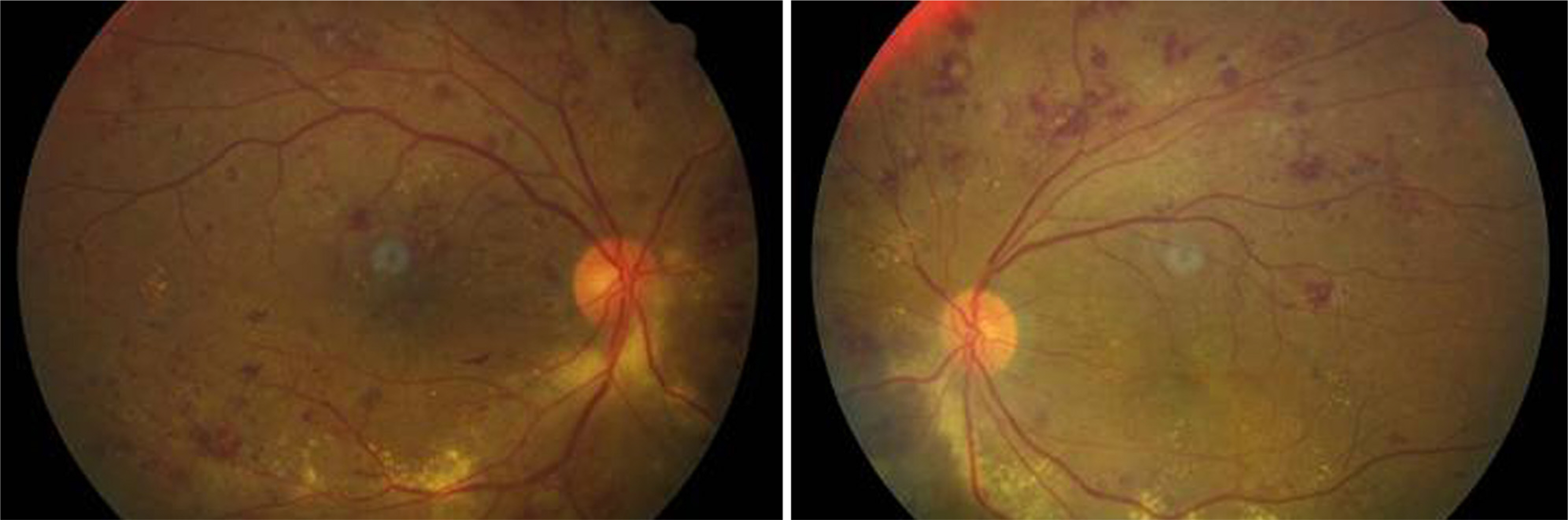
Figure 1. Fundus color photographs of the patient showed diffuse multiple dot and blot hemorrhages throughout the retina of both eyes.

Figure 2. Central macular OCT examinations showed retinal thickening with macular edema in both eyes.

Figure 3. Fluorescein angiogram showed diffuse microaneurysms and ischemia at the peripheral retina and posterior pole and leakage around the disc.
Reference
-
References
1. Schachat AP, Markowitz JA, Guyer DR, et al. Ophthalmic manifestations of leukaemia. Arch Ophthalmol. 1989; 107:697–700.2. Jampol LM, Goldberg MF, Busse B. Peripheral retinal microaneurysms in chronic leukemia. Am J Ophthalmol. 1975; 80:242–8.
Article3. Stephens DJ. Relation of viscosity of blood to leukocyte count, with particular reference to chronic myelogenous leukemia. Proc Soc Exp Biol Med. 1936; 35:251.4. Duke JR, Wilkinson CP, Sigelman S. Retinal microaneurysms in leukaemia. Br J Ophthalmol. 1968; 52:368–74.
Article5. Kincaid MC, Green WR. Ocular and orbital involvement in leukaemia. Surv Ophthalmol. 1983; 27:211–32.6. Rosenthal AR. Ocular manifestations of leukaemia. A review. Ophthalmology. 1983; 90:899–905.7. Morse PH, McCready JL. Peripheral retinal neovascularization in chronic myelocytic leukemia. Am J Ophthalmol. 1971; 72:975–8.
Article8. Frank RN, Ryan SJ Jr. Peripheral retinal neovascularization with chronic myelogenous leukemia. Arch Ophthalmol. 1972; 87:585–9.
Article9. Mandava N, Costakos D, Bartlett HM. Chronic myelogenous leukemia manifested as bilateral proliferative retinopathy. Arch Ophthalmol. 2005; 123:576–7.
Article10. Duane TD, Osher RH, Green WR. White centred haemorrhages: their significance. Ophthalmology. 1980; 87:66–9.11. Raynor MK, Clover A, Luff AJ. Leukaemia manifesting as uncon-trollable proliferative retinopathy in a diabetic. Eye. 2000; 14:400–1.
Article
- Full Text Links
-
- Actions
-
Cited
- CITED
-
- Close
- Share
-
- Similar articles
-
- Erythema Nodosum Associated with Chronic Myelogenous Leukemia
- Fundus Findings in Leukemia and Various Anemias
- Acute megakaryoblastic blast crisis as a presentation manifestation of chronic myelogenous leukemia
- Development of peripheral T-cell lymphoma in the course of chronic myelogenous leukemia
- Clinical Experience of Leukemia with Extradural Chloroma during Treatment of Lower Back Pain
