J Korean Ophthalmol Soc.
2009 Aug;50(8):1167-1173. 10.3341/jkos.2009.50.8.1167.
Tear Film and Ocular Surface Changes in Chronic Renal Failure Patients Undergoing Hemodialysis
- Affiliations
-
- 1Department of Ophthalmology, Chonnam National University Medical School, Gwangju, Korea. kcyoon@chonnam.ac.kr
- 2Department of Public Health, Graduate School Chonnam National University, Gwangju, Korea.
- KMID: 2212529
- DOI: http://doi.org/10.3341/jkos.2009.50.8.1167
Abstract
- PURPOSE
To evaluate the changes of tear film and ocular surface in patients with chronic renal failure (CRF) undergoing hemodialysis. METHODS: In 50 eyes of 25 CRF patients undergoing hemodialysis and 30 eyes of 15 control subjects, tear break-up time (BUT), basal tear secretion (BST), corneal sensation, tear clearance rate (TCR), keratoepitheliopathy and impression cytology findings were compared. Correlation between the above parameters and local or systemic factors was analyzed. RESULTS: Tear BUT, BST, TCR, conjunctival squamous metaplasia and conjunctival goblet cell density in the patient and control groups were 7.7+/-3.8 and 11.1+/-2.9 sec, 7.0+/-3.1 and 14.0+/-6.2 mm, 3.5+/-1.2 and 4.5+/-0.7, 1.5+/-0.6 and 0.3+/-0.5, 6.5+/-1.7 and 24.3+/-21.4 cells/mm2. Osmolarity in serum and tears of the patient group was higher than that of the control group. Serum and tear osmolarity correlated with BST, corneal sensation and conjunctival goblet cell density, and serum creatine level correlated with conjunctival goblet cell density (p<0.05). CONCLUSIONS: Patients with CRF undergoing hemodialysis have an increased risk of dry eye and thus, serum and tear osmolarity in dry eye patients should be closely monitored.
Keyword
MeSH Terms
Figure
-

Figure 1. Impression cytologic findings. (A) Specimen from a patient with chronic renal failure undergoing hemodialysis shows a loss of goblet cells and large, polygonal epithelial cells. (B) Specimen from a normal subject shows many periodic acid-Schiff positive goblet cells and small, round epithelial cells.
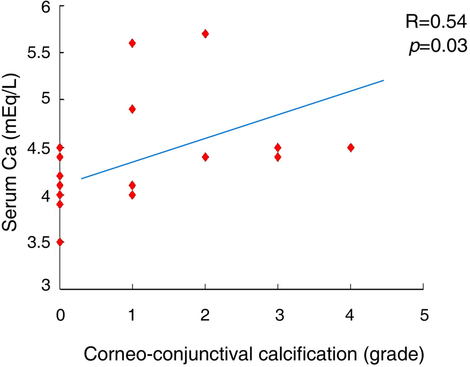
Figure 2. Correlation between corneoconjunctival calcification (grade) and serum calcium (mEq/L) in chronic renal failure patients undergoing hemodialysis.
Reference
-
References
1. Rechtman E, Harris A. An update on diabetic retinopathy. Harefuah. 2007; 146:872–7.2. Knox DL, Hanneken AM, Hollows FC, et al. Uremic optic neuropathy. Arch Ophthalmol. 1988; 106:50–4.
Article3. Klaassen-Broekema N, van Bijsterveld OP. Red eyes in renal failure. Br J Ophthalmol. 1992; 76:268–71.
Article4. Abrams JD. Corneal and other ocular findings in patients on intermittent dialysis for renal failure. Proc R Soc Med. 1966; 59:533–4.
Article5. Wang GL, Zhang F, Peng XY, Meng SM. The study of clinical characteristic of nonischemic central retinal vein occlusion. Zhonghua Yan Ke Za Zhi. 2008; 44:152–6.6. Brancati FL, Whelton PK, Randall BL, et al. Risk of end-stage renal disease in diabetes mellitus: a prospective cohort study of men screened for MRFIT. Multiple Risk Factor Intervention Trial. JAMA. 1997; 278:2069–74.
Article7. Comstock TJ. Renal dialysis. Young LY, Koda-Kimble MA, editors. Applied therapeutics: The clinical use of drugs. 6th ed.Vancouver: WA;1995. 31:chap. 1-15.8. Guy J, Johnston PK, Corbett JJ, et al. Treatment of visual loss in pseudotumor cerebri associated with uremia. Neurology. 1990; 40:28–32.
Article9. Walsh FB, Brown AB. Bilateral blindness of sudden onset. Trans Ophthalmol Soc. 1963; 23:13–27.10. Olivieri NF, Buncic RJ, Chew E, et al. Visual and auditory neurotoxicity in patients receiving subcutaneous desferrioxamine infusions. N Engl J Med. 1986; 314:869–73.11. Dursun D, Demirhan B, Oto S, Aydın P. Impression cytology of the conjunctival epithelium in patients with chronic renal failure. Br J Ophthalmol. 2000; 84:225–7.
Article12. Aktaş Z, Ozdek S, Asli Dinç U, et al. Alterations in ocular surface and corneal thickness in relation to metabolic control in patients with chronic renal failure. Nephrology. 2007; 12:380–5.13. Porter R, Crombie AL. Corneal and conjunctival calcification in chronic renal failure. Br J Ophthalmol. 1973; 57:339–43.
Article14. Ozdemir M, Bakaris S, Ozdemir G, et al. Ocular surface disorders and tear function changes in patients with chronic renal failure. Can J Ophthalmol. 2004; 39:526–32.15. Bakaris S, Ozdemir M, Isik IO, et al. Impression cytology changes and corneoconjunctival calcification in patients with chronic renal failure. Acta Cyto. 2005; 49:1–6.
Article16. Charlton JF, Schwab IR, Stuchell R. Tear hyperosmolarity in renal dialysis patients asymptomatic for dry eye. Cornea. 1996; 15:335–9.
Article17. Yoon KC, Im SK, Park YG, et al. Application of umbilical cord serum eyedrops for the treatment of dry eye syndrome. Cornea. 2006; 25:268–72.
Article18. Yoon KC, Heo H, Im SK, et al. Comparison of autologous serum and umbilical cord serum eye drops for dry eye syndrome. Am J Ophthalmol. 2007; 144:86–92.
Article19. Yoon KC, Jeong IY, Im SK, et al. Therapeutic effect of umbilical cord serum eyedrops for the treatment of dry eye associated with graft-versus-host disease. Bone Marrow Transplant. 2007; 39:231–5.
Article20. Yoon KC, Im SK, Seo MS. Changes of tear film and ocular surface in diabetes mellitus. Korean J Ophthalmol. 2004; 18:168–74.
Article21. Nelson JD. Impression cytology. Cornea. 1988; 7:71–81.
Article22. Inoue K, Kato S, Ohara C, et al. Ocular and systemic factors relevant to diabetic keratoepitheliopathy. Cornea. 2001; 20:798–801.
Article23. Tsubota K. Tear dynamics and dry eye. Prog Retin Eye Res. 1998; 17:565–96.
Article24. Kaido M, Goto E, Dogru M, Tsubota K. Punctal occlusion for Stevens-Johnson syndrome. Ophthalmology. 2004; 111:895–900.25. Schultz RO, Matsuda M, Yee RW, et al. Corneal endothelial changes in type 1 and type II diabetes mellitus. Am J Ophthalmol. 1984; 98:401–10.26. Herse PR. A review of manifestations of diabetes mellitus in the anterior eye and cornea. Am J Optom Physiol Optics. 1988; 65:224–30.
Article27. Hatchell DL, Magolan JJ Jr, Besson MJ, et al. Damage to the epithelial basement membrane in the corneas of diabetic rabbits. Arch Ophthalmol. 1983; 101:469–71.
Article28. Friend J, Thoft RA. The diabetic cornea. Int Ophthalmol Clin. 1984; 24:111–23.
Article29. Wong TY, Mitchell P. The eye in hypertension. Lancet. 2007; 369:425–35.
Article30. Schubert HD. Ocular manifestations of systemic hypertension. Curr Opin Ophthalmol. 1998; 9:69–72.
Article31. Vignanelli M, Stucchi CA. Conjunctival calcification in patients in chronic hemodialysis. Morphologic, clinical and epidemiologic study. J Fr Ophtalmol. 1988; 11:483–92.32. Demir N, Altay M, Ozer E, et al. Duration of renal failure as risk factor for conjunctival squamous metaplasia. Acta Cytol. 2008; 52:309–12.
Article33. The definition and classification of dry eye disease: report of the Definition and Classification Subcommittee of the International Dry Eye WorkShop (2007). Ocul Surf. 2007; 5:75–92.34. Seyahi N, Altiparmak MR, Kahveci A, et al. Association of conjunctival and corneal calcification with vascular calcification in dialysis patients. Am J Kidney Dis. 2005; 45:550–6.
Article35. Pahor D, Hojs R, Gracner B. Conjunctival and corneal changes in chronic renal failure patients treated with maintenance hemodialysis. Ophthalmologica. 1995; 209:14–6.
Article36. Oto S, Aydin P, Haberal A, et al. Is tear calcium an indicator of ocular calcification in patients with chronic renal failure? Eur J Ophthalmol. 1997; 7:181–4.
Article37. Tomlinson A, Khanal S, Ramaesh K, et al. Tear film osmolarity: determination of a referent for dry eye diagnosis. Invest Ophthalmol Vis Sci. 2006; 47:4309–15.
Article38. Gilbard JP, Farris RL, Santamaria J II. Osmolarity of tear micro-volumes in keratoconjunctivitis sicca. Arch Ophthalmol. 1978; 96:677–81.
Article39. Farris RL, Gilbard JP, Stutchell RN, Mandel ID. Diagnostic testsin keratoconjunctivitis sicca. CLAO J. 1990; 9:23–8.40. Ogasawara K, Mitsubayashi K, Tsuru T, Karube I. Electrical conductivity of tear fluid in healthy persons and keratoconjunctivitis sicca patients measured by a flexible conductimetric sensor. Graefes Arch Clin Exp Ophthalmol. 1996; 234:542–6.
Article41. Mastman GJ, Baldes EJ, Henderson JW. The total osmotic pressure of tears in normal and various pathologic conditions. Arch Ophthalmol. 1961; 65:509–13.
Article42. Broekema N, van Bijsterveld OP, de Bos Kuil RJ. Intraocular pressure during haemodialysis. Ophthalmologica. 1988; 197:60–4.
- Full Text Links
-
- Actions
-
Cited
- CITED
-
- Close
- Share
-
- Similar articles
-
- Clinical Experience of Hemodialysis on Three Cases of Renal Failure using Kill Type Artificial Kidney
- Recent treatment of dry eye
- Clinical Significance of Tear Film Osmolarity in Patients with Mild Dry Eye Syndrome
- Changes of Tear Film and Ocular Surface in Diabetes Mellitus
- Effects of Smoking on Tear Film and Ocular Surface
