Short-term Efficacy of Intravitreal Ranibizumab for Myopic Choroidal Neovascularization
- Affiliations
-
- 1Department of Ophthalmology and Visual Science, College of Medicine, The Catholic University of Korea, Seoul, Korea. youngjungroh@hanmail.net
- KMID: 2212490
- DOI: http://doi.org/10.3341/jkos.2009.50.7.1027
Abstract
- PURPOSE
To evaluate the effects of intravitreal ranibizumab in myopic choroidal neovascularization (CNV). METHODS: Patients who underwent intravitreal ranibizumab injection for myopic CNV, and were followed up more than 6 months, and their records were retrospectively investigated. The best corrected visual acuity, central macular thickness, and leak in fluorescein angiography were compared at baseline, and at 1, 3, and 6 months after injection. RESULTS: Twenty-one eyes of 18 patients were evaluated. The mean best corrected visual acuity (logMAR) was 1.23+/-0.65, 0.96+/- 0.40, 0.95+/-0.67, and 0.83+/-0.58 at baseline, 1, 3, and 6 months, respectively (p<0.001, p=0.006, p=0.001). The mean central macular thickness was 233.42+/-65.55 microm, 204.14+/-65.29 micrometer, and 157.76+/-71.45 microm at baseline, 3, and 6 months, respectively (p<0.001). In fluorescein angiography at 6 months after injection, regression was observed in 12 eyes, and fibrosis in 9 eyes. CONCLUSIONS: Intravitreal ranibizumab injection for myopic CNV in Korean patients appeared to be effective, resulting in regression of lesion and improvement of visual acuity.
MeSH Terms
Figure
-
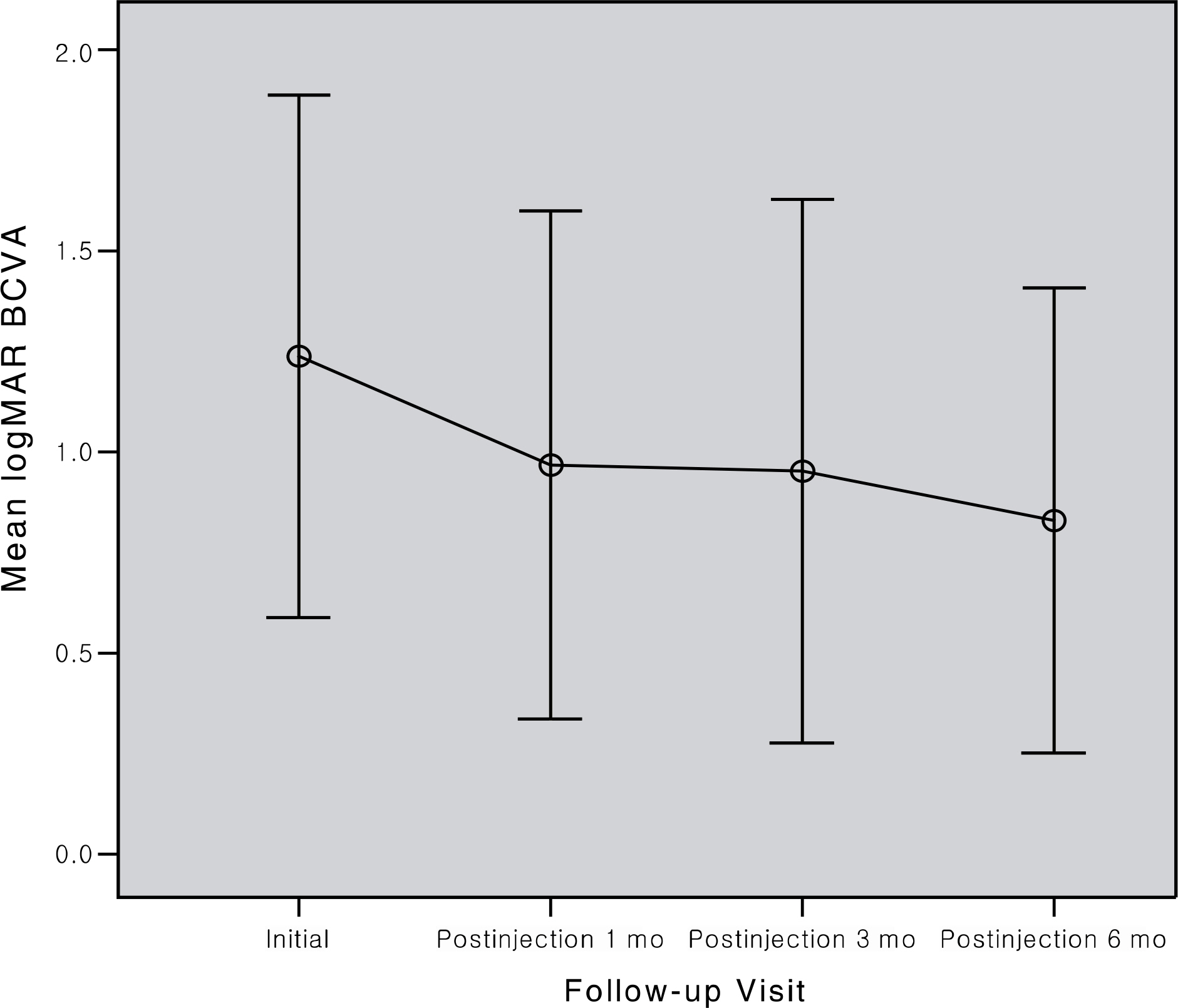
Figure 1. Graph showing changes in the mean loagarithm of the minimum angle of resolution (logMAR) best-corrected visual acuity (BCVA) after intravitreal ranibizumab treatment. Error bar, standard error of the mean.
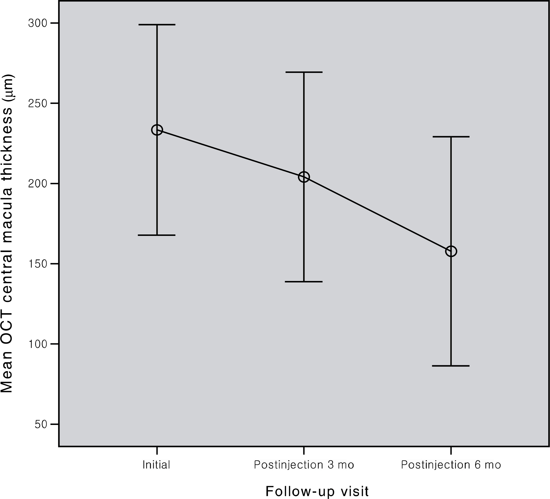
Figure 2. Graph showing changes in optical coherence tomography (OCT) central macular thickness after intravitreal ranibizumab treatment. Error bar, standard error of the mean.

Figure 3. The fundus photographs (A, B), fluorescein angiographs (FA, C-F), macula optical coherence tomographs (OCT, G, H) of patient 12. (A) fundus photograph before intravitreal ranibizumab injection showed subfoveal hemorrhage with chorioretinal atrophy. (B) At 6 months after intravitreal ranibizumab, fundus photograph showed resolution of juxtafoveal choroidal neovascularization (CNV). (C and E) arteriovenous and late venous phase FA before intravitreal ranibizumab injection showed leakage from juxtafoveal CNV. (D and F) arteriovenous and late venous phase FA at 6 months after intravitreal ranibizumab injection showed just stain due to retinal pigment epithelial atrophy. (G) OCT before treatment showed CNV. (H) OCT at 6 months after treatment showed resolution of CNV.
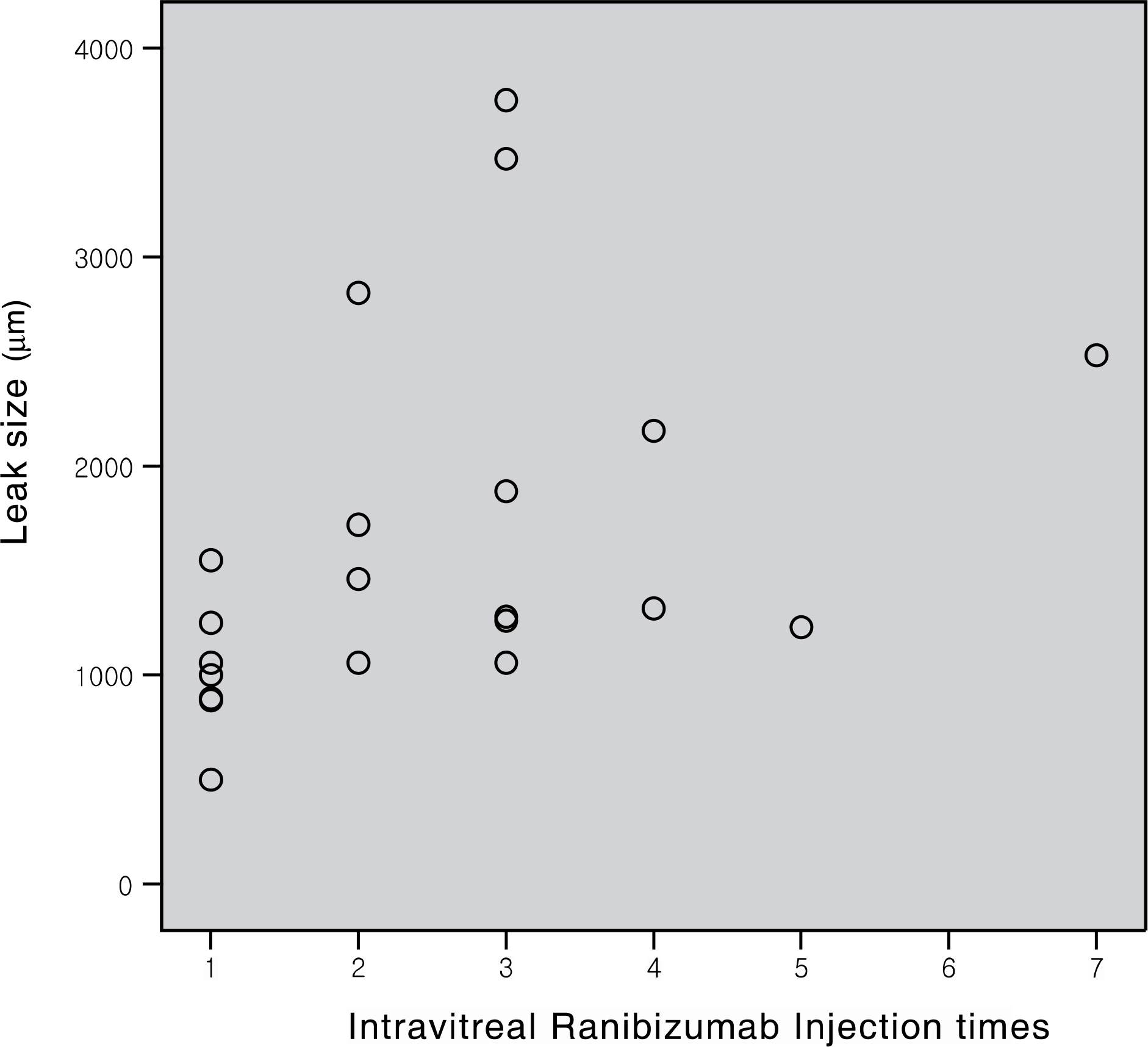
Figure 4. The relationship of the leak size and intravitreal ranibizumab injection times. The larger lesion needed more injection of intravitreal ranibizumab significantly (Spearman correlation coefficient rho=0.553, p=0.009).
Cited by 1 articles
-
Risk Factors for Retinal Pigment Epithelium Tears after Anti-VEGF Agent Injection in Age-Related Macular Degeneration
Woo Seok Choae, Jae Hong Park, Woo Seok Lee, Sang Won Kim, Hee Seong Yoon
J Korean Ophthalmol Soc. 2013;54(10):1546-1553. doi: 10.3341/jkos.2013.54.10.1546.
Reference
-
References
1. Grossniklaus HE, Green WR. Pathologic findings in pathologic myopia. Retina. 1992; 12:127–33.
Article2. Hotchkiss ML, Green WR. Pathologic myopia and choroidal neovascularization. Am J Ophthalmol. 1981; 91:177–83.
Article3. Tabandeh H, Flynn HW Jr, Scott IU, et al. Visual acuity outcomes of patients 50 years of age and older with high myopia and untreated choroidal neovascularization. Ophthalmology. 1999; 106:2063–7.
Article4. Yoshida T, Ohno-Matsui K, Ohtake Y, et al. Long-term visual prognosis of choroidal neovascularization in high myopia: a comparison between age groups. Ophthalmology. 2002; 109:712–9.5. Ryu IH, Kim BG, Lee SC. Photodynamic therapy of subfoveal choroidal neovascualrization in pathologic myopia. J Korean Ophthalmol Soc. 2003; 44:1991–5.6. Verteporfin in Photodynamic Therapy Study Group. Photo-dynamic therapy of subfoveal choroidal neovascularization in pathologic myopia with verteporfin: 1-year results of a randomized clinical trial—VIP report no. 1. Ophthalmology. 2001; 108:841–52.7. Blinder KJ, Blumenkranz MS, Bressler NM, et al. Verteporfin therapy of subfoveal choroidal neovascularisation in pathologic myopia: 2-year results of a randomized clinical trial—VIP report no. 3. Ophthalmology. 2003; 110:667–73.8. Emerson MV, Lauer AK, Flaxel CJ, et al. Intravitreal bevacizumab (Avastin) treatment of neovascular age-related macular degeneration. Retina. 2007; 27:439–44.
Article9. Goff MJ, Johnson RN, McDonald HR, et al. Intravitreal bevacizumab for previously treated choroidal neovascularization from age-related macular degeneration. Retina. 2007; 27:432–8.
Article10. Lazic R, Gabric N. Intravitreally administered bevacizumab (Avastin) in minimally classic and occult choroidal neovascularization secondary to age-related macular degeneration. Graefes Arch Clin Exp Ophthalmol. 2007; 245:68–73.
Article11. Costa RA, Jorge R, Calucci D, et al. Intravitreal Bevacizumab for choroidal neovascularization caused by AMD (IBeNA Study): results of a phase 1 dose-escalation study. Invest Ophthalmol Vis Sci. 2006; 47:4569–78.
Article12. Rich RM, Rosenfeld PJ, Puliafito CA, et al. Short-term safety and efficacy of intravitreal bevacizumab (Avastin) for neovascular agerelated macular degeneration. Retina. 2006; 26:495–511.
Article13. Hernandez-Rojas ML, Quiroz-Mercado H, Dalma-Weiszhausz J, et al. Short-term effects of intravitreal bevacizumab for subfoveal choroidal neovascularization in pathologic myopia. Retina. 2007; 27:707–12.14. Ruiz-Moreno JM, Gomez-Ulla F, Montero JA, et al. Intravitreous bevacizumab to treat subfoveal choroidal neovascularization in highly myopic eyes: short-term results. Eye. 2009; 23:334–8.
Article15. Brown DM, Kaiser PK, Michels M, et al. Ranibizumab versus verteporfin for neovascular age-related macular degeneration. N Engl J Med. 2006; 355:1432–44.
Article16. Silva RM, Ruiz-Moreno JM, Nascimento J, et al. Short-term efficacy and safety of intravitreal Ranibizumab for myopic choroidal neovascularization. Retina. 2008; 28:1117–23.
Article17. Brancato R, Pece A, Avanza P, Radrizzani E. Photocoagulation scar expansion after laser therapy for choroidal neovascularization in degenerative myopia. Retina. 1990; 10:239–43.
Article18. Ladas ID, Moschos MM, Rouvas AA, et al. Lacquer crack formation after photodynamic therapy. Eur J Ophthalmol. 2003; 13:729–33.
Article19. Ohno-Matsui K, Moriyama M, Hayashi K, Mochizuki M. Choroidal vein and artery occlusion following photodynamic therapy in eyes with pathologic myopia. Graefes Arch Clin Exp Ophthalmol. 2006; 244:1363–6.
Article20. Yamamoto I, Rogers AH, Reichel E, et al. Intravitreal bevacizumab (Avastin) as treatment for subfoveal choroidal neovascularisation secondary to pathological myopia. Br J Ophthalmol. 2007; 91:157–60.
Article21. Gharbiya M, Allievi F, Mazzeo L, Gabrieli CB. Intravitreal Bevacizumab Treatment for Choroidal Neovascularization in Pathologic Myopia: 12-month Results. Am J Ophthalmol. 2009; 147:84–93.
Article22. Chan WM, Lai TY, Liu DT, Lam DS. Intravitreal bevacizumab (Avastin) for myopic choroidal neovascularization: six-month results of a prospective pilot study. Ophthalmology. 2007; 114:2190–6.23. Ikuno Y, Sayanagi K, Soga K, et al. Intravitreal Bevacizumab for Choroidal Neovascularization Attributable to Pathological Myopia: One-Year Results. Am J Ophthalmol. 2009; 147:94–100.
Article24. Arias L, Planas N, Prades S, et al. Intravitreal bevacizuamb for choroidal neovascularisation secondary to pathological myopia: 6-month results. Br J Ophthalmol. 2008; 92:1035–9.25. Rosenfeld PJ, Rich RM, Lalwani GA. Ranibizumab: phase III clinical trial results. Ophthalmol Clin North Am. 2006; 19:361–72.26. Kojima A, Ohno-Matsui K, Teramukai S, et al. Estimation of visual outcome without treatment in patients with subfoveal choroidal neovascularization in pathologic myopia. Graefes Arch Clin Exp Ophthalmol. 2006; 244:1474–9.
Article27. Axer-Siegel R, Ehrlich R, Weinberger D, et al. Photodynamic therapy of subfoveal choroidal neovascularization in high myopia in a clinical setting: visual outcome in relation to age. Am J Ophthalmol. 2004; 138:602–7.
- Full Text Links
-
- Actions
-
Cited
- CITED
-
- Close
- Share
-
- Similar articles
-
- Long-term Therapeutic Effect of Intravitreal Bevacizumab (Avastin) on Myopic Choroidal Neovascularization
- Recurrent Idiopathic Choroidal Neovascularization in an Adolescent
- Long-Term Effect of Intravitreal Ranibizumab Injection on Choroidal Neovascularization in Age-Related Macular Degeneration
- Macular Hole Following Intravitreal Ranibizumab Injections for Choroidal Neovascularization
- Effect of High-dose Intravitreal Bevacizumab Injection on Refractory Idiopathic Choroidal Neovasculariz
