J Korean Ophthalmol Soc.
2009 Jun;50(6):887-892. 10.3341/jkos.2009.50.6.887.
Retinal Nerve Fiber Layer Thickness in Children With Glaucoma
- Affiliations
-
- 1Department of Ophthalmology, Oxford Eye Hospital, Headington, Oxford, UK.
- 2Nuffield Laboratory of Ophthalmology, University of Oxford, Oxford, UK.
- 3Advanced Medical and DentalInstitute, Universiti Sains Malaysia, Penang, Malaysia.
- 4Hangil Eye Hospital, Incheon, Korea.
- 5Department of Physiology, Anatomy and Genetics, University of Oxford, Oxford, UK.
- 6Department of Ophthalmology, College of Medicine, The Catholic University of Korea, Incheon St. Mary's Hospital, Incheon, Korea. yimhb@catholic.ac.kr
- KMID: 2212366
- DOI: http://doi.org/10.3341/jkos.2009.50.6.887
Abstract
-
PURPOSE: To find the optimal parameter of retinal nerve fiber layer (RNFL) analysis in optical coherence tomography (OCT) for diagnosing glaucoma in children.
METHODS
The study was comprised of 127 eyes of 84 patients (aged 6 to 18 years) who visited our institute between March 2006 and February 2008. Subjects were classified into normal, glaucoma suspect and glaucoma groups, and each eye was scanned using Stratus 3.0 OCT. Routine ophthalmic examinations including fundus examination, visual field test and OCT RNFL analysis were performed.
RESULTS
There were 55 normal eyes, 27 glaucoma suspect eyes and 45 glaucomatous eyes. The average RNFL thickness was the most useful parameter to differentiate between the glaucoma and non-glaucoma groups. The next most useful parameter was inferior average thickness, followed by superior RNFL thickness. The sensitivity and specificity of the new discriminant of the formula used were 78%, and 68.6%, respectively.
CONCLUSIONS
In OCT analysis, the average RNFL thickness is the most useful parameter in the diagnosis of glaucoma in children. The new discriminant of the formula is useful in the diagnosis of pediatric glaucoma patients.
MeSH Terms
Figure
-

Figure 1. Comparison of mean value between normal, glaucoma suspect and glaucomatous eye using optical coherence tomography retinal nerve fiber layer analysis.
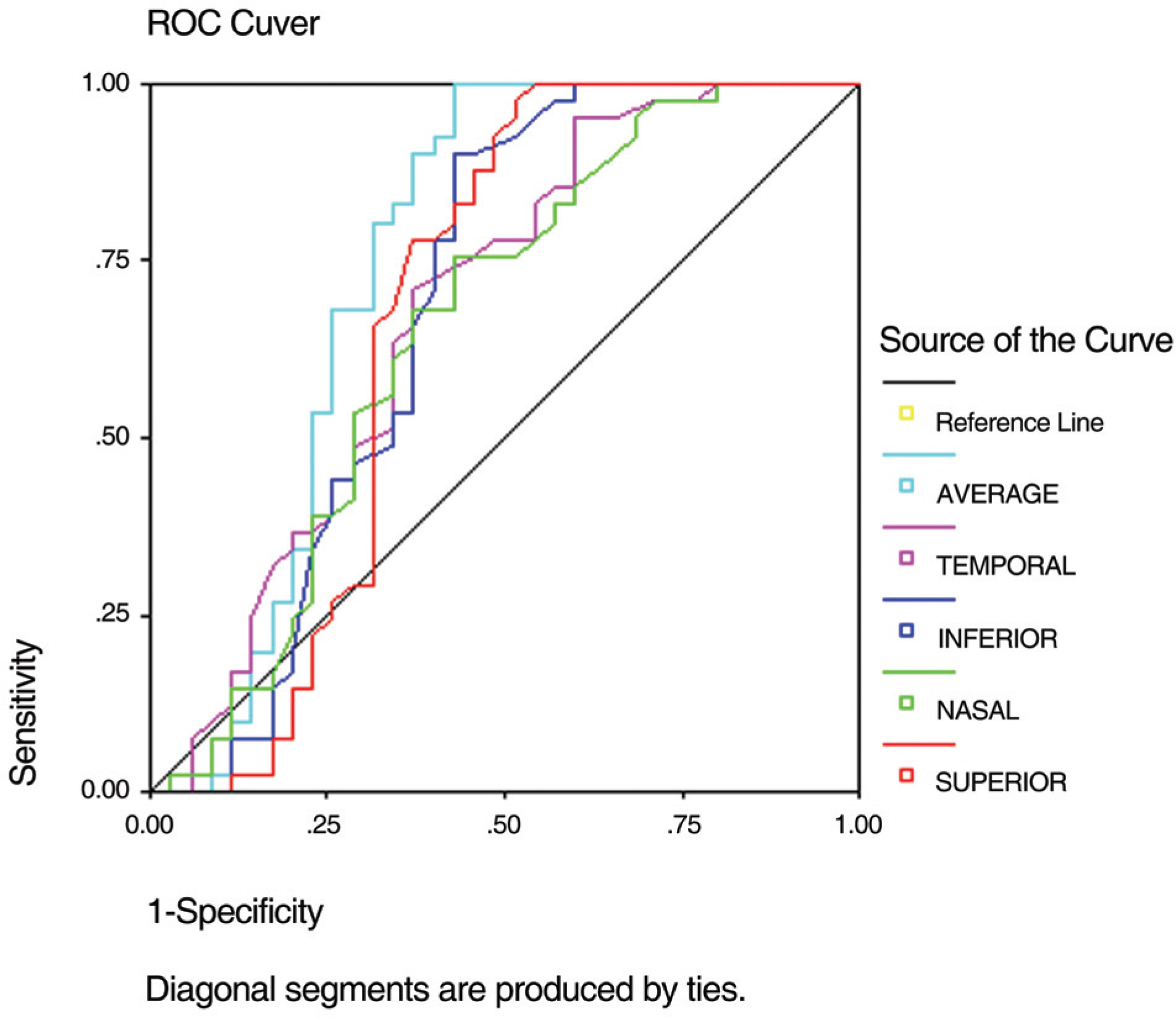
Figure 2. ROC (receiver operator characteristic) curve of the discriminant formula.
Cited by 1 articles
-
Retinal Nerve Fiber Layer Volume Measurements in Normal Children Using Spectral Domain Optical Coherence Tomography
Dong Eik Lee, Joong Won Shin, Han Woong Lim, Yong Un Shin, Min Ho Kang, Hee Yoon Cho, Min Cheol Seong
J Korean Ophthalmol Soc. 2016;57(5):800-807. doi: 10.3341/jkos.2016.57.5.800.
Reference
-
References
1. Quigley HA, Addicks EM, Green WR. Optic nerve damage in human glaucoma. Arch Ophthalmol. 1982; 100:135–46.
Article2. Quigley HA, Dunkelberger GR, Green WR. Retinal ganglion cell atrophy correlated with automated perimetry in human eyes with glaucoma. Am J Ophthalmol. 1989; 107:453–64.
Article3. Sommer A, Katz J, Quigley HA, et al. Clinically detectable nerve fiber atrophy precedes the onset of glaucomatous field loss. Arch Ophthalmol. 1991; 109:77–83.
Article4. Ahn HC, Son HW, Kim JS, Lee JH. Quantitative analysis of retinal nerve fiber layer thickness of normal children and adolescents. Korean J Ophthalmol. 2005; 19:195–200.
Article5. Ha SW, Rho SH. Age-related differences of optical coherence tomography data in Koreans. J Korean Ophthalmol Soc. 2005; 46:2037–44.6. Song JH, Kim ER, Yoo JM. Analysis of RNFL thickness and optic nerve head measured with OCT in children. J Korean Ophthalmol Soc. 2007; 48:1346–53.
Article7. Park SJ, Park KH, Yu YS, et al. Early detection of glaucoma with retinal nerve fiber layer photograph. J Korean Ophthalmol Soc. 1998; 39:180–6.8. Mrugacz M, Bakunowicz-Lazarczyk A. Optical coherence tomography measurement of the retinal nerve fiber layer in normal and juvenile glaucomatous eyes. Ophthalmologica. 2005; 219:80–5.
Article9. Varma R, Bazzaz S, Lai M. Optical tomography-measured retinal nerve fiber layer thickness in normal Latinos. Invest Ophthalmol Vis Sci. 2003; 44:3369–73.
Article10. Alamouti B, Funk J. Retinal thickness decreases with age: an OCT study. Br J Ophthalmol. 2003; 87:899–901.
Article11. Poinoosawmy D, Fontana L, Wu JX, et al. Variation of nerve fiber layer thickness measurements with age and ethnicity by scanning laser polarimetry. Br J Ophthalmol. 1997; 81:350–4.12. Kang KD, Park CK. Comparison of diagnostic precision between preprogramed indicator and newly calculated indicator in optical coherence tomography. J Korean Ophthalmol Soc. 2006; 47:243–52.13. Cho YK, Lee YC, Lee SY. Factors mediating effects on the retinal nerve fiber layer thickness in normal children. J Korean Ophthalmol Soc. 2008; 49:98–103.
Article14. Hwang YH, Kang JH. Artifacts in retinal nerve fiber layer analysis using optical coherence tomography. J Korean Ophthalmol Soc. 2008; 49:778–83.
Article15. Bowd C, Zangwill LM, Blumenthal EZ, et al. Imaging of the optic disc and retinal nerve fiber layer: The effects of age, optic disc area, refractive error, and gender. J Opt Soc Am A Opt Image Sci Vis. 2002; 19:197–207.
Article
- Full Text Links
-
- Actions
-
Cited
- CITED
-
- Close
- Share
-
- Similar articles
-
- Reproducibility of Retinal Nerve Fiber Layer Thickness Evaluation by Nerve Fiber Analyzer
- Biometry of Retinal Nerve Fiber Layer Thickness by NFA
- Influence of Diabetes Mellitus on the Retinal Ne rve Fiber Layer Thickness Measurement by Nerve Fiber Analyzer
- Clinical Evaluation of Unilateral Open-Angle Glaucoma: A Two-Year Follow-Up Study
- Differentiating Patients with Glaucoma from Glaucoma Suspects by Retinal Nerve Fiber Layer Assessment Using Nerve Fiber Analyzer
