J Korean Ophthalmol Soc.
2009 May;50(5):774-778. 10.3341/jkos.2009.50.5.774.
Corneal Melting and Descemetocele Resulting From Noninfectious Keratitis Related to the Cosmetic Contact Lenses
- Affiliations
-
- 1Department of Ophthalmology, HanGil Eye Hospital, Incheon, Korea. chobjn@empal.com
- KMID: 2212323
- DOI: http://doi.org/10.3341/jkos.2009.50.5.774
Abstract
-
PURPOSE:To report 2 cases of corneal melting and corneal melting with descemetocele that occurred in users of cosmetic contact lenses.
CASE SUMMARY
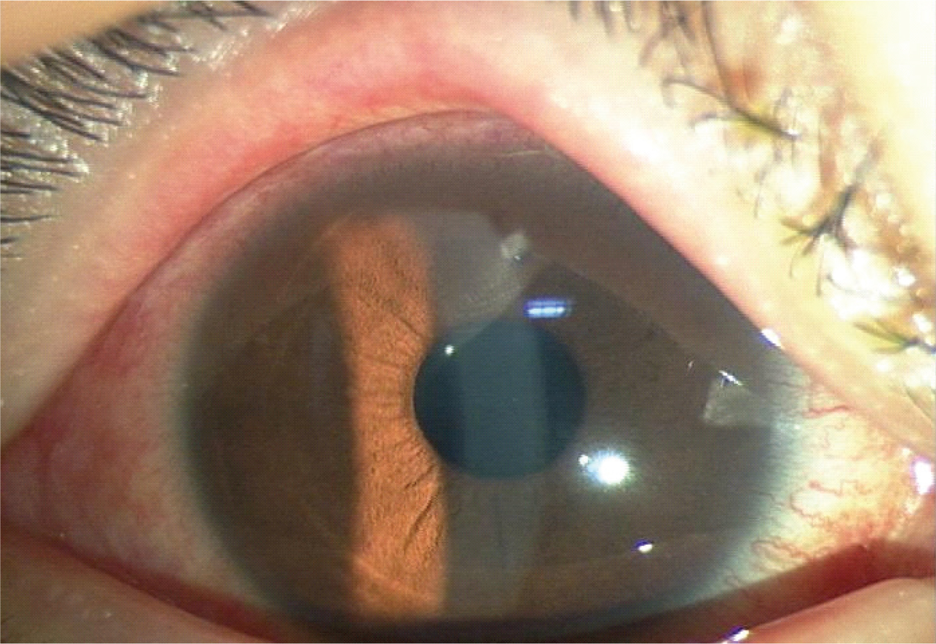
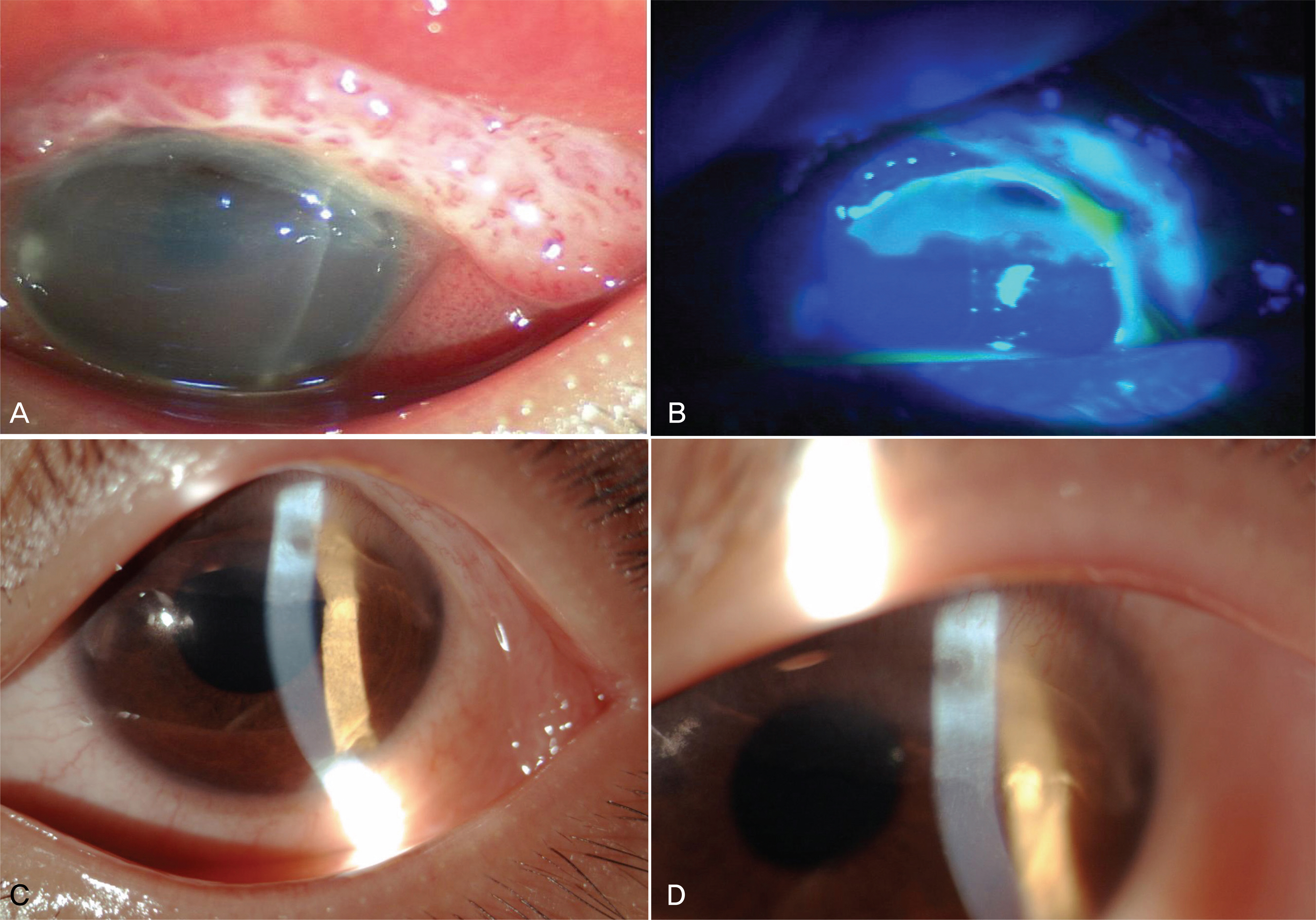
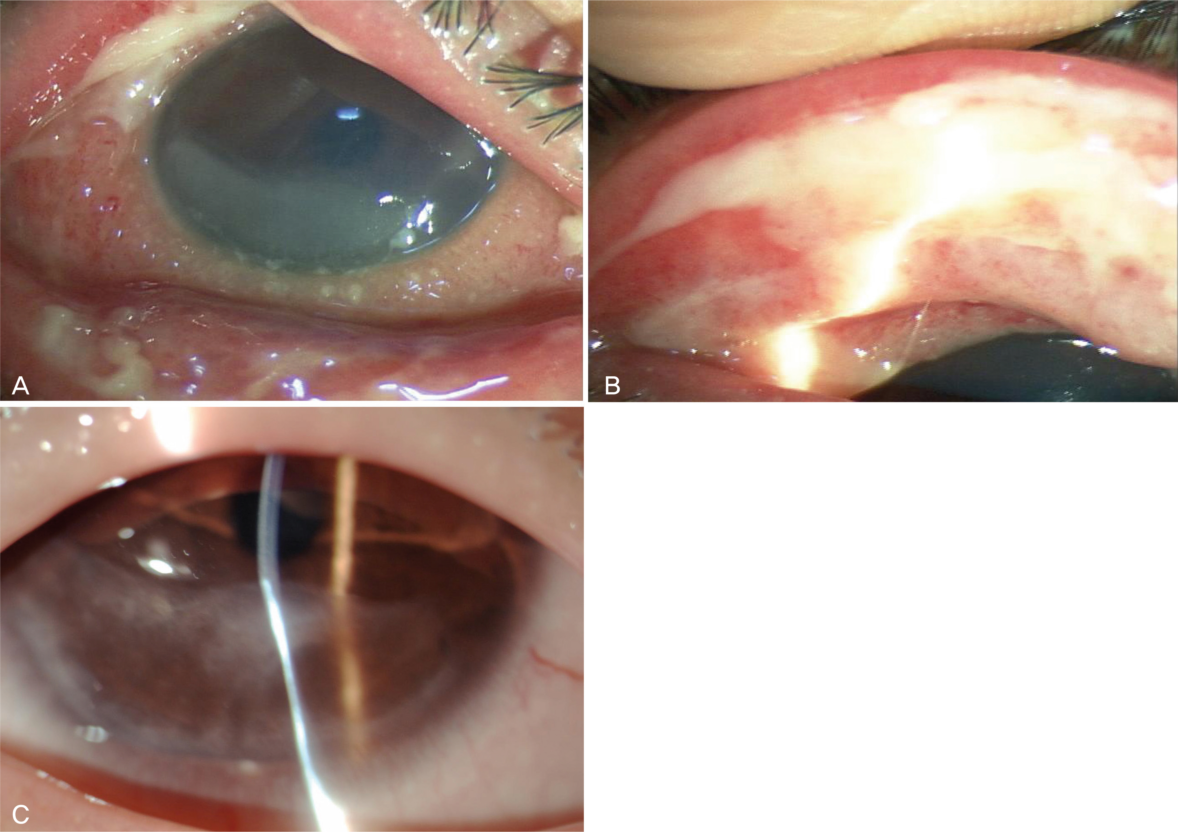
A-12-year-old and a 13-year-old female who used cosmetic contact lenses were referred to our clinic under the preliminary diagnosis of keratitis and corneal melting. The patients had purchased the lenses from an optician and had worn the lenses for approximately 1 month without being educated on their proper use. The signs and symptoms improved after 2 weeks of treatment with oral steroid and 1% topical prednisolone acetate. However, descemetocele occurred in the 12-year-old patient. Reepithelization of the cornea had been completed within the treatment period. However, corneal thinning with mild opacity remained in the lesions, and the best corrected visual acuities on the Snellen chart were 20/30 in both patients.
MeSH Terms
Figure
Cited by 1 articles
-
Comparison of Surface Roughness and Bacterial Adhesion between Cosmetic Contact Lenses and Conventional Contact Lenses
Yong Woo Ji, Soon Ho Hong, Dong Yong Chung, Eung Kweon Kim, Hyung Keun Lee
J Korean Ophthalmol Soc. 2014;55(5):646-655. doi: 10.3341/jkos.2014.55.5.646.
Reference
-
References
1. Lee WJ, Yoon GS, Shyn KH. Corneal Complication in Contact Lens Wearer. J Korean Ophthalmol Soc. 1996; 37:225–32.2. Park YM, Hahn TW, Choi SH, et al. Acanthamoeba Keratitis Related to Cosmetic Contact Lenses. J Korean Ophthalmol Soc. 2007; 48:991–4.3. Efron N, Morgan PB, Hill EA, et al. The size, location and clinical severity of corneal infiltrative events associated with contact lens wear. Optom Vis Sci. 2005; 82:519–27.
Article4. Donshik PC, Sucheki JK, Ehlers WH. Peripheral corneal infiltrates associated with contact lens wear. Trans Am Ophthalmol Soc. 1995; 93:49–60.5. Stein RM, Clinch TE, Cohen FJ, et al. Infected vs sterile corneal infiltrates in contact lens wearers. Am J Ophthalmol. 1988; 105:632–6.
Article6. Bates AK, Morris RJ, Stapleton F. Sterile corneal infiltrates in contact lens wearers. Eye. 1989; 3:803–10.
Article7. Sweeney DF, Jalbert I, Covey M, et al. Clinical characterization of corneal infiltrative events observed with soft contact lens wear. Cornea. 2003; 22:435–42.
Article8. Steinemann TL, Pinninti U, Szczotka LB, et al. Ocular complication associated with the use of cosmetic contact lenses from unlicensed vendors. Eye Contact Lens. 2003; 29:196–200.
- Full Text Links
- Actions
-
Cited
- CITED
-
- Close
- Share
- Similar articles
-
- Infectious Keratitis Associated with Contact Lenses
- Characteristics and Complications of Cosmetic Contact Lens
- Acanthamoeba Keratitis Related to Cosmetic Contact Lenses
- A Case of Fungal Keratitis as a Complication of Orthokeratology Contact Lens
- Keratitis with Elizabethkingia meningoseptica Occurring after Contact Lens Wear: A Case Report


