Changes in the Cornea and Anterior Chamber After LASEK: Pentacam Findings
- Affiliations
-
- 1Department of Ophthalmology, KyungHee University Medical Center, Seoul, Korea. khjinmd@khmc.or.kr
- 2Department of Ophthalmology, KyungHee University East-west Neo Medical Center, Seoul, Korea.
- KMID: 2212250
- DOI: http://doi.org/10.3341/jkos.2009.50.4.510
Abstract
-
PURPOSE: We evaluated changes in the cornea and anterior chamber after LASEK using Pentacam to search for signs of subclinical keratectasia occurrence.
METHODS
Seventy-one eyes of 36 patients who had received LASEK were enrolled in this study. All eyes were examined for asphericity of the anterior and posterior cornea (Q-value), anterior and posterior corneal displacement, central corneal thickness, anterior chamber depth, angle, and volume using Pentacam before surgery and again one month after the operation.
RESULTS
The postoperative changes in the asphericity of the posterior cornea and posterior corneal displacement were not statistically significant(p=0.668, p=0.101). The anterior chamber depth, angle, and volume decreased by 0.088 mm, 0.983degrees, 7.21 mm3 after LASEK, respectively. (p=0.000) The postoperative changes in asphericity of the anterior cornea, anterior corneal displacement, and central corneal thickness were statistically significant(p=0.000).
CONCLUSIONS
In this study, we compared changes in the anterior chamber and cornea after LASEK using Pentacam. Changes in the anterior cornea were significant; however, the posterior cornea did not change significantly. The anterior chamber depth, anterior chamber angle, and volume were decreased one month after the operation, although further long-term follow-ups will be necessary to verify these findings.
Keyword
MeSH Terms
Figure
-

Figure 1. The scatterplots for corneal asphericity between preoperative and postoperative states (at 1 month after operation). (A) Anterior corneal surface. A prolate anterior corneal surface had changed into an oblate surface after operation. (B) Posterior corneal surface. An asphericity of the posterior cornea was unchanged after operation.
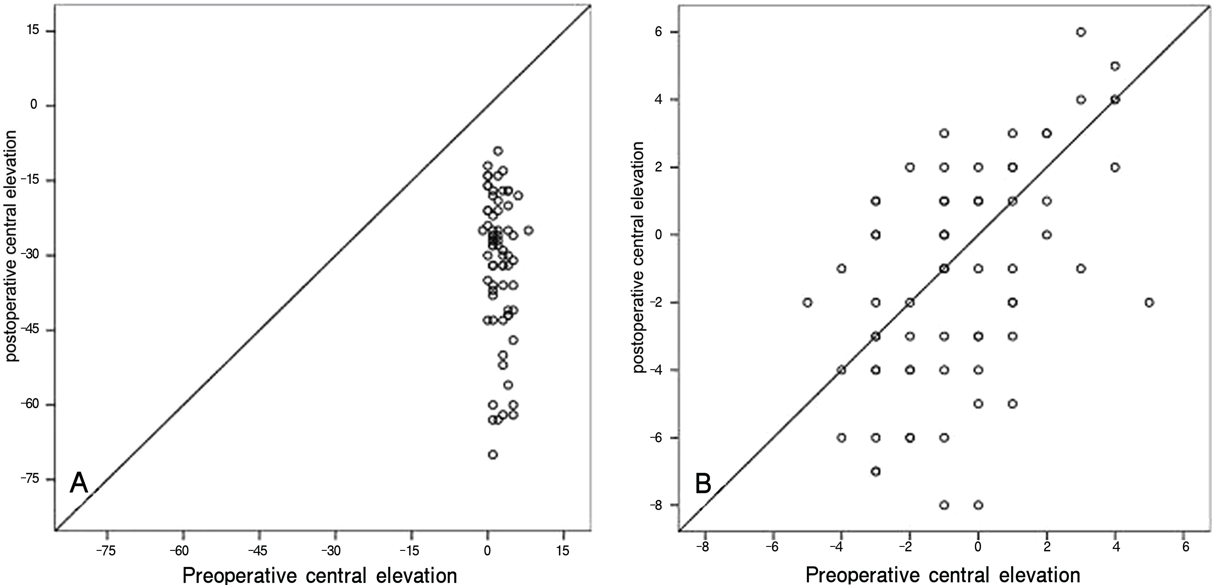
Figure 2. The scatterplots for corneal central elevation between preoperative and postoperative states (at 1 month after operation). (A) Anterior corneal surface. Central elevation of anterior corneal surface was lower after operation.(B) Posterior corneal surface. Central elevation of posterior corneal surface was unchanged after operation.

Figure 3. The scatterplots for anterior chamber parameters between preoperative and postoperative states (at 1 month after operation). (A) Anterior chamber depth. Anterior chamber depth had become shallow after operation. (B) Anterior chamber angle. Anterior chamber angle had become narrow after operation. (C) Anterior chamber volume. Anterior chamber volume had shrunk after operation.

Figure 4. The schematic figures of change of the cornea after LASEK surgery. (A) Section of cornea during LASEK surgery. (B) Magnification of section plane and free body diagram. (C) Magnification of section plane and net force of free body diagram. Net force of free body diagram was directed posterior to anterior through axial plane. (D) Change of cornea and anterior chamber after LASEK surgery. Posterior cornea was not displaced and the anterior cornea was displaced posteriorly. Because of tensile strength, the iris was displaced anteriorly resulting in the anterior chamber angle, volume, and depth was decreased.
Cited by 1 articles
-
Clinical Results of Wavefront-guided LASIK
Jung Taeck Hong, Jooeun Lee, Jae Yong Kim, Myoung Joon Kim, Hungwon Tchah
J Korean Ophthalmol Soc. 2010;51(11):1438-1444. doi: 10.3341/jkos.2010.51.11.1438.
Reference
-
References
1. Kim ES, Jin KH. Evaluation of prophylactic use of mitomycin to inhibit haze formation after LASEK. J Korean Ophthalmol Soc. 2007; 48:623–9.2. Swartz T, Marten L, Wang M. Measuring the cornea: the latest developments in corneal topography. Curr Opin Ophthalmol. 2007; 18:325–33.
Article3. Rabsiber TM, Khoramnia R, Auffarth GU. Anterior chamber measurement using Pentacam rotating Scheimpflug camera. J Cataract Refract Surg. 2006; 32:456–9.4. Nemeth G, Vajas A, Kolozsvari B, et al. Anterior chamber depth measurements in phakic and pseudophakic eyes: Pentacam versus ultrasound device. J Cataract Refract Surg. 2006; 32:1331–5.
Article5. Buehl W, Stojanac D, Sacu S, et al. Comparison of three methods of measuring corneal thickness and anterior chamber depth. Am J Ophthalmol. 2006; 141:7–12.
Article6. Reuland MS, Reuland AJ, Nishi Y, Auffarth GU. Corneal radii and anterior chamber depth measurments using the IOLmaster versus the Pentacam. J Refract Surg. 2007; 23:368–73.7. Elbaz U, Barkana Y, Gerber Y, et al. Comparison of different techniques of anterior chamber depth and keratometric measurements. Am J Ophthalmol. 2007; 143:48–53.
Article8. Shanker H, Taranath D, Santhirathelagan CT, Pesudovs K. Anterior segment biometry with the Pentacam: Comprehensive assessment of repeatability of automated measurements. J Cataract Refract Surg. 2008; 34:103–13.9. Chen D, Lam AK. Intrasession and intersession repeatability of the Pentacam system on posterior corneal assessement in the normal human eye. J Cataract Refract Surg. 2007; 33:448–54.10. Sanctis UD, Loiacono C, Richiardi L, et al. Sensitivity and specificity of posterior corneal elevation measured by Pentacam in discriminating keratoconus/subclinical keratoconus. Ophthalmology. 2008; 9:1–6.
Article11. Ho JD, Tsai CY, Tsai RJ, et al. Validity of keratometric index: evaluation by the Pentacam rotating Scheimpflug camera. J Cataract Refract Surg. 2008; 34:137–45.12. Emre S, Doganay S, Yologlu S. Evaluation of anterior segment parameters in keratoconic eyes measured with the Pentacam system. J Cataract Refract Surg. 2007; 33:1708–12.
Article13. Anera RG, Jiménez JR, del Barci LJ, et al. Changes in corneal asphericity after laser in situe keratomileusis. J Cataract Refract Surg. 2003; 29:762–8.14. Ciolino JB, Khachikian SS, Cortese MJ, Belin MW. Long-term stability of the posterior cornea after laser in situ keratomileusis. J Cataract Refract Surg. 2007; 33:1366–70.
Article15. Hashemi H, Mehravaran S. Corneal changes after laser refractive surgery for myopia: comparison of Orbscan II and Pentacam findings. J Cataract Refract Surg. 2007; 33:841–7.
Article16. Calossi A. Corneal asphericity and spherical aberration. J Refract Surg. 2007; 23:505–14.
Article17. Xu L, Cao WF, Wang YX, et al. Anterior chamber depth and chamber angle and their associations with ocular and general parameters: the Beijing eye study. Am J Ophthalmol. 2008; 145:929–36.
Article18. Tamburrelli C, Giudiceandrea A, Vaiano AS, et al. Underestimate of tonometric reading after photorefractive keratectomy increases at higher intraocular pressure levels. Invest Ophthalmol Vis Sci. 2005; 46:3208–13.19. Svedberg H, Chen E, Hamberg-Nyström H. Changes in corneal thickness and curvature after different excimer laser photo-refractive procedure and their impact on intraocular pressure measurements. Graefes Arch Clin Exp Ophthalmol. 2005; 243:1218–20.20. Tsukiyama J, Miyamoto Y, Higaki S, et al. Changes in the anterior and posterior radii of the corneal curvature and anterior chamber depth by orthokeratology. Eye Contact Lens. 2008; 34:17–20.
Article21. Emre S, Çankaya C, Demirel S, Doganay S. Comparison of preoperative and postoperative anterior segment measurements with Pentacam in horizontal muscle surgery. Eur J Ophthalmol. 2008; 18:7–12.
Article22. Wong TY, Klein BE, Klein R, et al. Refractive errors, intraocular pressure, and glaucoma in a white population. Ophthalmology. 2003; 110:211–7.
- Full Text Links
-
- Actions
-
Cited
- CITED
-
- Close
- Share
-
- Similar articles
-
- Comparison of Corneal Measurement Values between Two Types of Topography
- The Changes in the Cornea and Anterior Chamber after Lateral Rectus Muscle Recession in Intermittent Extropia
- Comparison of Corneal Thickness and Anterior Chamber Depth Measured With Orbscan, Pentacam, and Ultrasound Pachymetry
- The Changes of Corneal High-Order Aberrations and Anterior Chamber Parameters after Trabeculectomy Using Pentacam(R)
- Measurement Comparison of Anterior Segment Parameters between AL-Scan(R) and Pentacam(R)
