J Korean Ophthalmol Soc.
2014 Jun;55(6):801-808.
Measurement Comparison of Anterior Segment Parameters between AL-Scan(R) and Pentacam(R)
- Affiliations
-
- 1Department of Ophthalmology, Inje University Busan Paik Hospital, Inje University College of Medicine, Busan, Korea.
- 2Shinsegae Eye Clinic, Busan, Korea. happytriad@gmail.com
Abstract
- PURPOSE
To investigate the clinical availability of AL-Scan(R) (Nidek, GAMAGORI, Japan) by comparing anterior segment parameters measured with AL-Scan(R) and Pentacam(R) (Oculus, Wetzlar, Germany).
METHODS
Seventy-three patients (117 eyes) who received refractive surgery at our hospital were tested with AL-Scan(R) and Pentacam(R). We compared measurements including anterior chamber depth, central corneal thickness, white-to-white, and corneal curvature.
RESULTS
When comparing measurements obtained with AL-Scan(R) and Pentacam(R), the anterior chamber depth (p < 0.001), central corneal thickness (p < 0.001) and 2.4 mm zone K value (p = 0.038) showed significant differences; the white-to-white (p = 0.348) and 3.3 mm zone K value (p = 0.429) showed no significant differences. All AL-Scan(R) and Pentacam(R) parameters had a strong positive linear correlation (p < 0.001). The Bland-Altman plots showed a high degree of agreement between AL-Scan(R) and Pentacam(R) in all parameters except for anterior chamber depth.
CONCLUSIONS
AL-Scan(R) is convenient to use clinically because simultaneous measurements of ocular biometry including axial length, intraocular lens power, and topography are possible. However, because differences in some anterior segment parameters exist when compared with Pentacam(R), measurements with AL-Scan(R) may require comparisons with other instruments.
Keyword
Figure
-
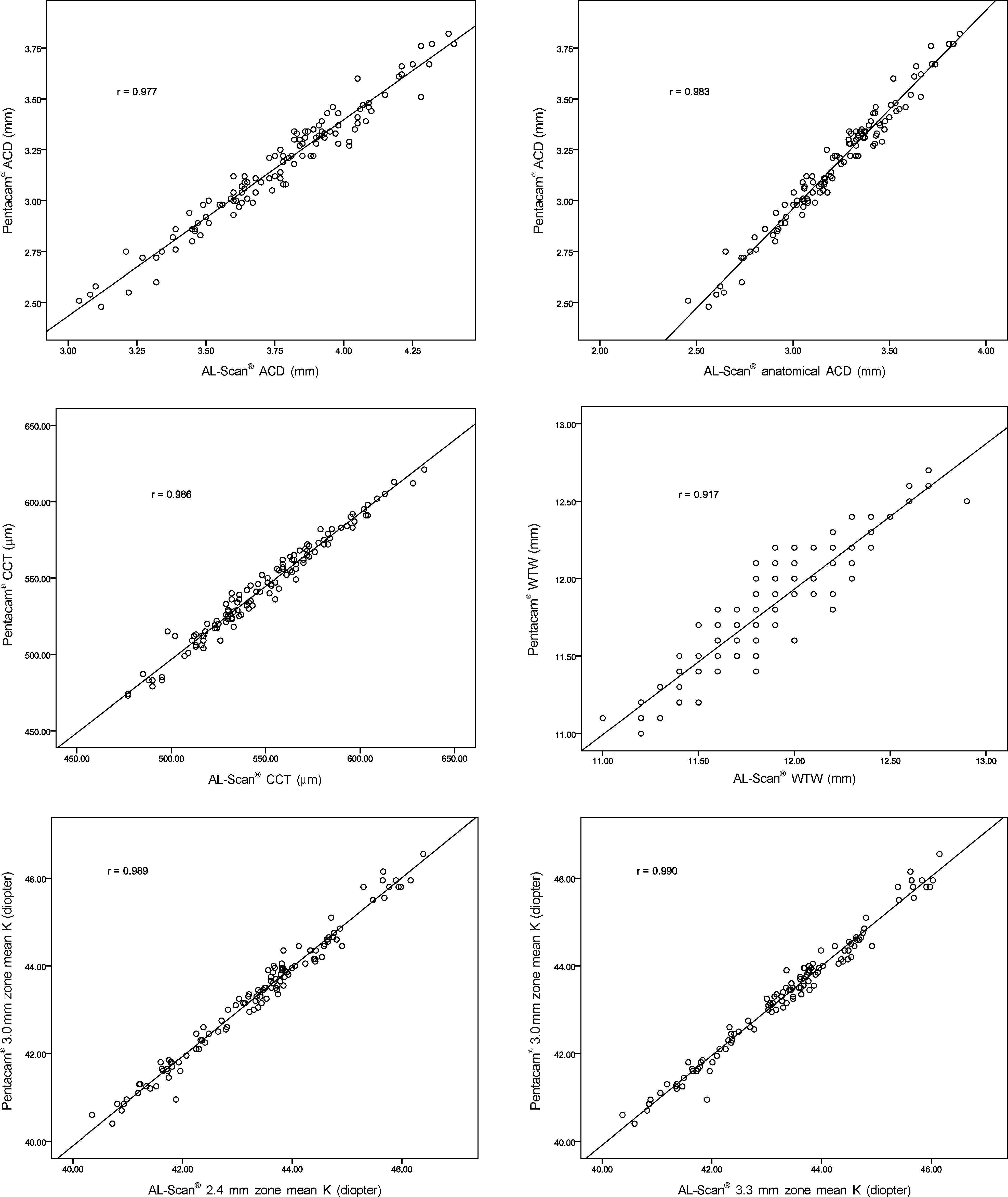
Figure 1. Pearson correlation of AL-Scan® and Pentcam® in anterior segment parameters. ACD = anterior chamber depth; CCT = central corneal thickness; WTW = white-to-white; K = keratometric diopter.

Figure 2. Bland-Altman plots of AL-Scan® and Pentacam® in anterior segment parameters. ACD = anterior chamber depth; CCT = central corneal thickness; WTW= white-to-white; K = keratometric diopter.
Reference
-
References
1. Kim DW, Yi KY, Choi DG, Shin YJ. Corneal thickness measured by dual Scheimpflug, anterior segment optical coherence tomog-raphy, and ultrasound pachymetry. J Korean Ophthal Soc. 2012; 53:1412–8.
Article2. Rüfer F, Schröder A, Arvani MK, Erb C. Central and peripheral cor-neal pachymetry–standard evaluation with the Pentacam system. Klin Monbl Augenheilkd. 2005; 222:117–22.3. Lee DM, Ahn JM, Seo KY, et al. Comparison of corneal measure-ment values between two types of topography. J Korean Ophthalmol Soc. 2012; 53:1584–90.
Article4. Shankar H, Taranath D, Santhirathelagan CT, Pesudovs K. Anterior segment biometry with the Pentacam: comprehensive assessment of repeatability of automated measurements. J Cataract Refract Surg. 2008; 34:103–13.
Article5. Kawamorita T, Nakayama N, Uozato H. Repeatability and reprodu-cibility of corneal curvature measurements using the Pentacam and Keratron topography systems. J Refract Surg. 2009; 25:539–44.
Article6. Menassa N, Kaufmann C, Goggin M, et al. Comparison and re-producibility of corneal thickness and curvature readings obtained by the Galilei and the Orbscan II analysis systems. J Cataract Refract Surg. 2008; 34:1742–7.
Article7. Li H, Leung CK, Wong L, et al. Comparative study of central cor-neal thickness measurement with slit-lamp optical coherence to-mography and visante optical coherence tomography. Ophthalmology. 2008; 115:796–801.
Article8. Savini G, Carbonelli M, Barboni P, Hoffer KJ. Repeatability of au-tomatic measurements performed by a dual Scheimpflug analyzer in unoperated and post-refractive surgery eyes. J Cataract Refract Surg. 2011; 37:302–9.
Article9. Savini G, Barboni P, Carbonelli M, Hoffer KJ. Repeatability of au-tomatic measurements by a new Scheimpflug camera combined with Placido topography. J Cataract Refract Surg. 2011; 37:1809–16.
Article10. Rosa N, Lanza M, Borrelli M, et al. Comparison of central corneal thickness measured with Orbscan and Pentacam. J Refract Surg. 2007; 23:895–9.
Article11. Kim SI, Kang SJ, Oh TH, et al. Accuracy of ocular biometry and postoperative refraction in cataract patients with AL-Scan®. J Korean Ophthalmol Soc. 2013; 54:1688–93.12. Buehl W, Stojanac D, Sacu S, et al. Comparison of three methods of measuring corneal thickness and anterior chamber depth. Am J Ophthalmol. 2006; 141:7–12.
Article13. Shin YJ, Kim NH, Kim DH. Comparison of Pentacam with Orbscan. J Korean Ophthalmol Soc. 2007; 48:637–41.14. Park JY, Kim SY, Jung MS. Comparison of corneal thickness and anterior chamber depth measured with Orbscan, Pentacam, and ultrasound pachymetry. J Korean Ophthalmol Soc. 2009; 50:664–9.
Article15. Ryu HW, Kim KR, Chung SK. Comparison of A-scan, Scheimpflug camera, and Orbscan for measurement of anterior chamber depth. J Korean Ophthalmol Soc. 2006; 47:1287–91.16. Kwag JY, Choi SH. Comparison of ocular biometry measured by ultrasound and two kinds of partial coherence interferometers. J Korean Ophthalmol Soc. 2011; 52:169–74.
Article17. Park Y, Hwang HB, Chung SK. Comparison of anterior chamber depth obtained from applanation and optical principle devices. J Korean Ophthalmol Soc. 2013; 54:1219–26.
Article18. Savini G, Carbonelli M, Sbreglia A, et al. Comparison of anterior segment measurements by 3 Scheimpflug tomographers and 1 Placido corneal topographer. J Cataract Refract Surg. 2011; 37:1679–85.
Article19. Amano S, Honda N, Amano Y, et al. Comparison of central corneal thickness measurements by rotating Scheimpflug camera, ultra-sonic pachymetry, and scanning-slit corneal topography. Ophthalmology. 2006; 113:937–41.
Article20. O'Donnell C, Maldonado-Codina C. Agreement and repeatability of central thickness measurement in normal corneas using ultrasound pachymetry and the OCULUS Pentacam. Cornea. 2005; 24:920–4.21. Lackner B, Schmidinger G, Pieh S, et al. Repeatability and reprodu-cibility of central corneal thickness measurement with Pentacam, Orbscan, and ultrasound. Optom Vis Sci. 2005; 82:892–9.
Article22. Kawamorita T, Uozato H, Kamiya K, et al. Repeatability, reprodu-cibility, and agreement characteristics of rotating Scheimpflug photography and scanning-slit corneal topography for corneal power measurement. J Cataract Refract Surg. 2009; 35:127–33.
Article23. Jain R, Dilraj G, Grewal SP. Repeatability of corneal parameters with Pentacam after laser in situ keratomileusis. Indian J Ophthalmol. 2007; 55:341–7.
- Full Text Links
-
- Actions
-
Cited
- CITED
-
- Close
- Share
-
- Similar articles
-
- Comparative Analysis of Corneal Refractive Power Measured with AL-Scan(R), Autokeratometer, and Pentacam(R)
- Comparision of Anterior Segment Parameters in Angle-Closure Glaucoma Using Scheimpflug Camera
- Comparison of Measurement of Anterior Segment Parameters Between Scheimpflug Camera and Ultrasound Biomicroscopy
- Anterior Segment Parameters Using Pentacam and Prediction of Corneal Endothelial Cell Loss after Cataract Surgery
- Comparison of Anterior Chamber Depth Obtained from Applanation and Optical Principle Devices
