Assessment of buccal bone thickness of aesthetic maxillary region: a cone-beam computed tomography study
- Affiliations
-
- 1Research Centre in Dental Sciences (CICO), Universidad de La Frontera Dental School, Temuco, Chile. ramon.fuentes@ufrontera.cl
- 2Department of Dental Materials and Prosthodontics, University of Sao Paulo Dental School of Ribeirao Preto, Ribeirao Preto, Brazil.
- KMID: 2212004
- DOI: http://doi.org/10.5051/jpis.2015.45.5.162
Abstract
- PURPOSE
The aim of this study was to analyze the anatomical dimensions of the buccal bone walls of the aesthetic maxillary region for immediate implant placement, based upon cone-beam computed tomography (CBCT) scans in a sample of adult patients.
METHODS
Two calibrated examiners analyzed a sample of 50 CBCT scans, performing morphometric analyses of both incisors and canines on the left and right sides. Subsequently, in the sagittal view, a line was traced through the major axis of the selected tooth. Then, a second line (E) was traced from the buccal to the palatal wall at the level of the observed bone ridges. The heights of the buccal and palatal bone ridges were determined at the major axis of the tooth. The buccal bone thickness was measured across five lines. The first was at the level of line E. The second was at the most apical point of the tooth, and the other three lines were equidistant between the apical and the cervical lines, and parallel to them. Statistical analysis was performed with a significance level of P< or =0.05 for the bone thickness means and standard deviations per tooth and patient for the five lines at varying depths.
RESULTS
The means of the buccal wall thicknesses in the central incisors, lateral incisors and canines were 1.14+/-0.65 mm, 0.95+/-0.67 mm and 1.15+/-0.68 mm, respectively. Additionally, only on the left side were significant differences in some measurements of buccal bone thickness observed according to age and gender. However, age and gender did not show significant differences in heights between the palatal and buccal plates. In a few cases, the buccal wall had a greater height than the palatal wall.
CONCLUSIONS
Less than 10% of sites showed more than a 2-mm thickness of the buccal bone wall, with the exception of the central incisor region, wherein 14.4% of cases were > or =2 mm.
MeSH Terms
Figure
-
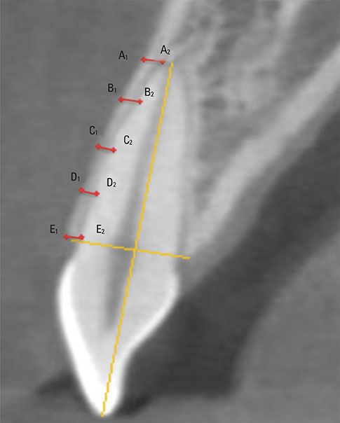
Figure 1 Analyses of buccal bone wall thicknesses in a sagittal cut of a CBCT scan at the labelled parallel lines. Line A1-A2 was located in the most apical tooth portion; Line E1-E2 was located at the level of both bone crests; Lines B, C and D were distributed equidistantly and parallel to each other in the space between lines A1-A2 and E1-E2.

Figure 2 Confidence intervals (95%) for buccal bone wall thicknesses on each line and tooth according to age range and side. *Significant differences (ANOVA).
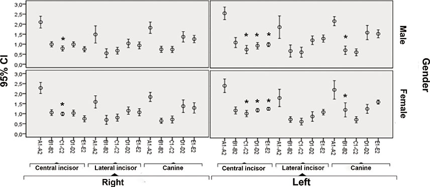
Figure 3 Confidence intervals (95%) for buccal bone wall thicknesses on each line and tooth according to gender and side. *Significant differences (ANOVA).
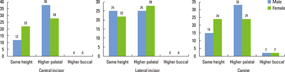
Figure 4 Classifications in height of bone ridges at each tooth according to gender. Note that the buccal bone wall never was higher than the palatal at both incisors.
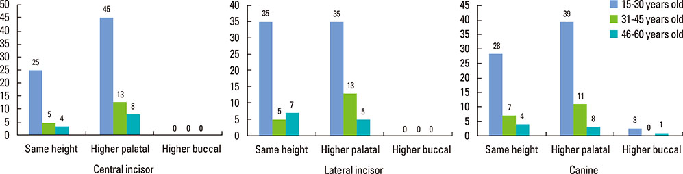
Figure 5 Classification of the height of the bone ridges at each tooth distributed by age range.
Cited by 2 articles
-
Cone-beam computed tomographic analysis of the alveolar ridge profile and virtual implant placement for the anterior maxilla
Hyun-Chang Lim, Do-Uk Kang, Hyehyeon Baek, Ji-Youn Hong, Seung-Yun Shin, Jong-Hyuk Chung, Yeek Herr, Seung-Il Shin
J Periodontal Implant Sci. 2019;49(5):299-309. doi: 10.5051/jpis.2019.49.5.299.Three-dimensional observations of the incisive foramen on cone-beam computed tomography image analysis
Yeon-Tae Kim, Jae-Hong Lee, Seong-Nyum Jeong
J Periodontal Implant Sci. 2020;50(1):48-55. doi: 10.5051/jpis.2020.50.1.48.
Reference
-
1. Buser D, Martin W, Belser UC. Optimizing esthetics for implant restorations in the anterior maxilla: anatomic and surgical considerations. Int J Oral Maxillofac Implants. 2004; 19:43–61.2. Rodriguez AM, Rosenstiel SF. Esthetic considerations related to bone and soft tissue maintenance and development around dental implants: report of the Committee on Research in Fixed Prosthodontics of the American Academy of Fixed Prosthodontics. J Prosthet Dent. 2012; 108:259–267.
Article3. Kim HJ, Yu SK, Lee MH, Lee HJ, Kim HJ, Chung CH. Cortical and cancellous bone thickness on the anterior region of alveolar bone in Korean: a study of dentate human cadavers. J Adv Prosthodont. 2012; 4:146–152.
Article4. Caubet Biayna J, Heras Rincón I, Sánchez Mayoral J, Morey Mas M, Iriarte Ortabe JI. Manejo de defectos óseos anteroposteriores en el frente estético. Rev Esp Cir Oral Maxilofac. 2009; 31:81–97.
Article5. Araújo MG, Sukekava F, Wennström JL, Lindhe J. Tissue modeling following implant placement in fresh extraction sockets. Clin Oral Implants Res. 2006; 17:615–624.
Article6. Chen ST, Darby IB, Reynolds EC. A prospective clinical study of non-submerged immediate implants: clinical outcomes and esthetic results. Clin Oral Implants Res. 2007; 18:552–562.
Article7. Engelke W, Beltrán V, Fuentes R, Decco O. Endoscopically Assisted Root Splitting (EARS): Method and First Results. Int J Odontostomatol. 2012; 6:313–316.
Article8. Beltrán V, Matthijs A, Borie E, Fuentes R, Valdivia-Gandur I, Engelke W. Bone healing in transverse maxillary defects with different surgical procedures using anorganic bovine bone in humans. Int J Morphol. 2013; 31:75–81.
Article9. Evans CD, Chen ST. Esthetic outcomes of immediate implant placements. Clin Oral Implants Res. 2008; 19:73–80.
Article10. Huynh-Ba G, Pjetursson BE, Sanz M, Cecchinato D, Ferrus J, Lindhe J, et al. Analysis of the socket bone wall dimensions in the upper maxilla in relation to immediate implant placement. Clin Oral Implants Res. 2010; 21:37–42.
Article11. Botticelli D, Berglundh T, Lindhe J. Hard-tissue alterations following immediate implant placement in extraction sites. J Clin Periodontol. 2004; 31:820–828.
Article12. Han JY, Jung GU. Labial and lingual/palatal bone thickness of maxillary and mandibular anteriors in human cadavers in Koreans. J Periodontal Implant Sci. 2011; 41:60–66.
Article13. Januário AL, Duarte WR, Barriviera M, Mesti JC, Araújo MG, Lindhe J. Dimension of the facial bone wall in the anterior maxilla: a cone-beam computed tomography study. Clin Oral Implants Res. 2011; 22:1168–1171.
Article14. Nowzari H, Molayem S, Chiu CH, Rich SK. Cone beam computed tomographic measurement of maxillary central incisors to determine prevalence of facial alveolar bone width ≥2 mm. Clin Implant Dent Relat Res. 2012; 14:595–602.
Article15. Lee SL, Kim HJ, Son MK, Chung CH. Anthropometric analysis of maxillary anterior buccal bone of Korean adults using cone-beam CT. J Adv Prosthodont. 2010; 2:92–96.
Article16. González-Martín O, Oteo C, Ortega R, Alandez J, Sanz M, Veltri M. Evaluation of peri-implant buccal bone by computed tomography: an experimental study. Clin Oral Implants Res. Forthcoming 2015.
Article17. Alqerban A, Jacobs R, Fieuws S, Willems G. Comparison of two cone beam computed tomographic systems versus panoramic imaging for localization of impacted maxillary canines and detection of root resorption. Eur J Orthod. 2011; 33:93–102.
Article18. Vera C, De Kok IJ, Reinhold D, Limpiphipatanakorn P, Yap AK, Tyndall D, et al. Evaluation of buccal alveolar bone dimension of maxillary anterior and premolar teeth: a cone beam computed tomography investigation. Int J Oral Maxillofac Implants. 2012; 27:1514–1519.19. Braut V, Bornstein MM, Lauber R, Buser D. Bone dimensions in the posterior mandible: a retrospective radiographic study using cone beam computed tomography. Part 1--analysis of dentate sites. Int J Periodontics Restorative Dent. 2012; 32:175–184.20. Johnson K. A study of the dimensional changes occurring in the maxilla following tooth extraction. Aust Dent J. 1969; 14:241–244.
Article21. Araújo MG, Lindhe J. Dimensional ridge alterations following tooth extraction. An experimental study in the dog. J Clin Periodontol. 2005; 32:212–218.
Article22. Schropp L, Wenzel A, Kostopoulos L, Karring T. Bone healing and soft tissue contour changes following single-tooth extraction: a clinical and radiographic 12-month prospective study. Int J Periodontics Restorative Dent. 2003; 23:313–323.23. Tomasi C, Sanz M, Cecchinato D, Pjetursson B, Ferrus J, Lang NP, et al. Bone dimensional variations at implants placed in fresh extraction sockets: a multilevel multivariate analysis. Clin Oral Implants Res. 2010; 21:30–36.
Article24. Sanz M, Cecchinato D, Ferrus J, Pjetursson EB, Lang NP, Lindhe J. A prospective, randomized-controlled clinical trial to evaluate bone preservation using implants with different geometry placed into extraction sockets in the maxilla. Clin Oral Implants Res. 2010; 21:13–21.
Article25. Ghassemian M, Nowzari H, Lajolo C, Verdugo F, Pirronti T, D'Addona A. The thickness of facial alveolar bone overlying healthy maxillary anterior teeth. J Periodontol. 2012; 83:187–197.
Article26. Belser U, Martin W, Jung R, Hämmerle C, Schmid B, Morton D, et al. ITI Treatment Guide, Volume 1. Implant therapy in the esthetic zone: single-tooth replacements. 1st ed. Berlin: Quintessence Publishing Co. Ltd.;2007.27. Grunder U, Gracis S, Capelli M. Influence of the 3-D bone-to-implant relationship on esthetics. Int J Periodontics Restorative Dent. 2005; 25:113–119.28. Qahash M, Susin C, Polimeni G, Hall J, Wikesjo UM. Bone healing dynamics at buccal peri-implant sites. Clin Oral Implants Res. 2008; 19:166–172.
Article29. Younes F, Eghbali A, Raes M, De Bruyckere T, Cosyn J, De Bruyn H. Relationship between buccal bone and gingival thickness revisited using non-invasive registration methods. Clin Oral Implants Res. Forthcoming 2015.
Article30. Nevins M, Camelo M, De Paoli S, Friedland B, Schenk RK, Parma-Benfenati S, et al. A study of the fate of the buccal wall of extraction sockets of teeth with prominent roots. Int J Periodontics Restorative Dent. 2006; 26:19–29.
Article31. Zekry A, Wang R, Chau AC, Lang NP. Facial alveolar bone wall width - a cone-beam computed tomography study in Asians. Clin Oral Implants Res. 2014; 25:194–206.
Article
- Full Text Links
-
- Actions
-
Cited
- CITED
-
- Close
- Share
-
- Similar articles
-
- Assessment of the relationship between the maxillary molars and adjacent structures using cone beam computed tomography
- Analysis of the root position and angulation of maxillary premolars in alveolar bone using cone-beam computed tomography
- Analysis of the root position of the maxillary incisors in the alveolar bone using cone-beam computed tomography
- A cone-beam computed tomography evaluation of buccal bone thickness following maxillary expansion
- Detection of maxillary second molar with two palatal roots using cone beam computed tomography: a case report
