J Korean Ophthalmol Soc.
2008 May;49(5):771-777. 10.3341/jkos.2008.49.5.771.
The Relationship Between Parameters Measured by Optical Coherence Tomography and Visual Field Indices
- Affiliations
-
- 1Department of Ophthalmology, Hanyang University College of Medicine, Guri Hospital, Gyeonggi, Korea.
- 2Department of Ophthalmology, Ulsan University, College of Medicine, Asan Medical Center, Seoul, Korea. mskook@amc.seoul.kr
- 3Hangil Eye Hospital, Incheon, Korea.
- 4Kangnam BS Eye Center, Seoul, Korea.
- KMID: 2211707
- DOI: http://doi.org/10.3341/jkos.2008.49.5.771
Abstract
-
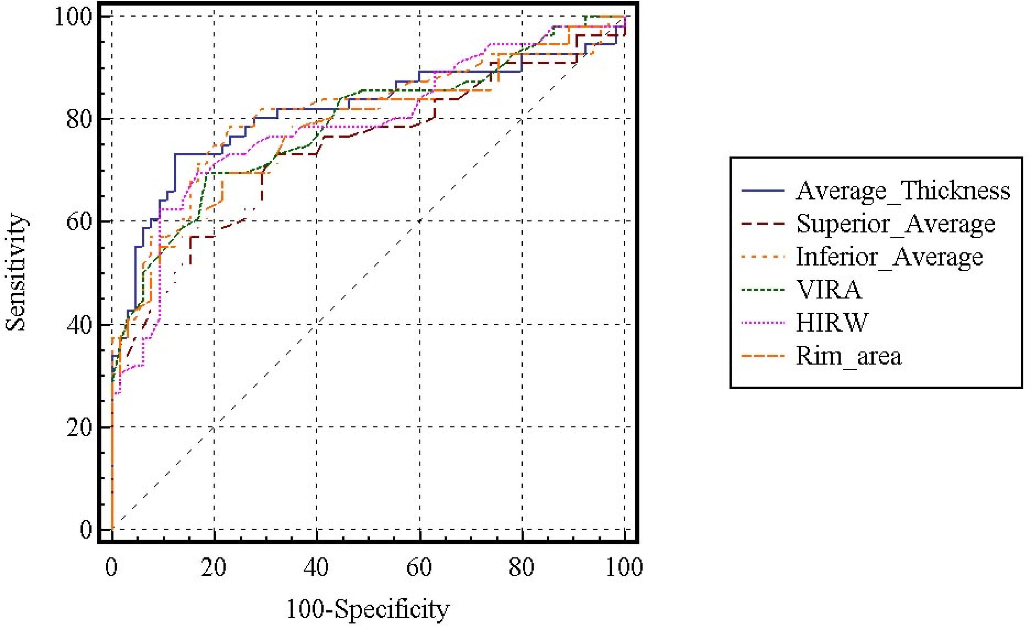
PURPOSE: To evaluate the diagnostic ability of optic disc topographic parameters and the retinal nerve fiber layer (RNFL) thickness parameter measured by optical coherence tomography (OCT) and to determine the association of these structural parameters with visual field indices.
METHODS
Fifty-six glaucomatous eyes and 65 healthy control eyes were enrolled in this retrospective cross-sectional study. Each subject had a 24-2 full threshold test on a Humphrey visual field analyzer and an optical coherence tomographic evaluation. The parameters from the fast RNFL thickness algorithm and the fast optic disc algorithm were analyzed by an ROC curve, and we sought to determine the association of these parameters with visual field indices by linear and logarithmic regression.
RESULTS
The area under the receiver operating characteristic curve (AUROC) value of the fast optic disc algorithm parameters ranged from 0.78 to 0.79 and that of the fast RNFL thickness algorithm parameters ranged from 0.74 to 0.81. The associations between the parameters from the fast optic disc algorithm and from the fast RNFL thickness algorithm with visual field indices were statistically significant (P<0.001).
CONCLUSIONS
The fast optic disc algorithm and the fast RNFL algorithm revealed comparable diagnostic ability in discriminating glaucoma and significant associations with visual field indices.
Keyword
MeSH Terms
Figure
Cited by 3 articles
-
Development of an LCD-Based Visual Field System
Jin-Ho Joo, Jihyoung Lee, Heecheon You, Jaheon Kang
J Korean Med Sci. 2018;33(3):. doi: 10.3346/jkms.2018.33.e19.Changes in Visual Field Index After Cataract Extraction
Sohee Jeon, Jeong Hoon Choi, Ji Wook Yang, Young Chun Lee, Su Young Kim
J Korean Ophthalmol Soc. 2010;51(3):386-392. doi: 10.3341/jkos.2010.51.3.386.The Stereoscopic Acuity in Patients with Unilateral or Bilateral Visual Field Defects
Joo Hyun Chang, Bo Young Chun, Jae Pil Shin
J Korean Ophthalmol Soc. 2014;55(5):734-739. doi: 10.3341/jkos.2014.55.5.734.
Reference
-
References
1. Sommer A, Miller NR, Pollack I, et al. The nerve fiber layer in the diagnosis of glaucoma. Arch Ophthalmol. 1977; 95:2149–56.
Article2. Zangwill LM, Williams J, Berry CC, et al. A comparison of optical coherence tomography and retinal nerve fiber layer photography for detection of nerve fiber layer damage in glaucoma. Ophthalmology. 2000; 107:1309–15.
Article3. Kim JM, Park KH, Kim TW, Kim DM. Comparison of the results between Heldelberg Retina Tomograph II and Stratus Optical Coherence Tomography in Glaucoma. J Korean Ophthalmol Soc. 2006; 47:556–62.4. Sim JO, Park CK. Optic nerve head analysis obtained by optical coherence tomography for the diagnosis of glaucoma in Koreans. J Korean Ophthalmol Soc. 2004; 45:1885–92.5. Manassakorn A, Nouri-Mahdavi K, Caprioli J. Comparison of retinal nerve fiber layer thickness and optic disk algorithms with optical coherence tomography to detect glaucoma. Am J Ophthalmol. 2006; 141:105–15.
Article6. Garway-Heath DF, Holder GE, Fitzke FW, Hitchings RA. Relationship between electrophysiological, psychophysical, and anatomical measurements in glaucoma. Invest Ophthalmol Vis Sci. 2002; 43:2213–20.7. Schlottmann PG, De Cilla S, Greenfield DS, et al. Relationship between visual field sensitivity and retinal nerve fiber layer thickness as measured by scanning laser polarimetry. Invest Ophthalmol Vis Sci. 2004; 45:1823–9.
Article8. Leung CK, Chong KK, Chan WM, et al. Comparative study of retinal nerve fiber layer measurement by StratusOCT and GDx VCC, II: structure/function regression analysis in glaucoma. Invest Ophthalmol Vis Sci. 2005; 46:3702–11.
Article9. Reus NJ, Lemij HG. Relationships between standard automated perimetry, HRT confocal scanning laser ophthalmoscopy, and GDx VCC scanning laser polarimetry. Invest Ophthalmol Vis Sci. 2005; 46:4182–8.
Article10. Zangwill LM, Bowd C, Berry CC, et al. Discriminating between normal and glaucomatous eyes using the Heidelberg retinal tomography, GDx nerve fiber analyzer, and optical coherence tomograph. Arch Ophthalmol. 2001; 119:985–93.11. Bowd C, Sangwill LM, Berry CC, et al. Detecting early glaucoma by assessment of retinal nerve fiber layer thickness and visual function. Invest Ophthalmol Vis Sci. 2001; 42:1993–2003.12. Kanamori A, Nakamura M, Escano MF, et al. Evaluation of the glaucomatous damage on retinal nerve fiber layer thickness measured by optical coherence tomography. Am J Ophthalmol. 2003; 135:513–20.
Article13. Medeiros FA, Zangwill LM, Bowd C, et al. Evaluation of retinal nerve fiber layer, optic nerve head, and macular thickness measurements for glaucoma detection using optical coherence tomography. Am J Ophthalmol. 2005; 139:44–55.
Article14. Schuman JS, Tamar PK, Hertzmark E, et al. Reproducibility of nerve fiber layer thickness measurements using optical coherence tomography. Ophthalmology. 1996; 103:1889–98.
Article15. Schuman JS, Hee MR, Arya AB, et al. Optical coherence tomography: a new tool for glaucoma diagnosis. Curr Opin Ophthalmol. 1995; 6:89–95.
Article16. Wang M, Luo R, Liu Y. Optical coherence tomography and its application in ophthalmology. Yan Ke Xue Bao. 1998; 14:116–20.17. Baumal CR. Clinical applications of optical coherence tomography. Curr Opin Ophthalmol. 1999; 10:182–8.
Article18. Wollstein G, Ishikawa H, Wang J, et al. Comparison of three optical coherence tomography scanning areas for detection of glaucomatous damage. Am J Ophthalmol. 2005; 139:39–43.
Article19. Lai E, Wollstein G, Price LL, et al. Optical coherence tomography disc assessment in optic nerves with peripapillary atrophy. Ophthalmic Surg Lasers Imaging. 2003; 34:498–504.20. Schuman JS, Wollstein G, Farra T, et al. Comparison of optic nerve head measurements obtained by optical coherence tomography and confocal scanning laser ophthalmoscopy. Am J Ophthalmol. 2003; 135:504–12.
Article21. Quigley HA, Katz J, Derrick RJ, et al. An evaluation of optic disc and nerve fiber layer examinations in monitoring progression of early glaucoma damage. Ophthalmology. 1992; 99:19–28.
Article22. Sommer HA, Quigley HA, Robin AL, et al. Evaluation of nerve fiber layer assessment. Arch Ophthalmol. 1984; 102:1766–71.
Article23. Tuulonen A, Airaksinen PJ. Initial glaucomatous optic disc and retinal nerve fiber layer abnormalities and their progression. Am J Ophthalmol. 1991; 111:485–90.
- Full Text Links
- Actions
-
Cited
- CITED
-
- Close
- Share
- Similar articles
-
- Influence of Epiretinal Membranes on the Retinal Nerve Fiber Layer Thickness Measured by Spectral Domain Optical Coherence Tomography in Glaucoma
- Correlation between Heidelberg Retina Tomograph, Visual Field, and Optical Coherence Tomography in Primary Open-Angle Glaucoma
- Correlation Between Central Corneal Thickness and Glaucomatous Damage
- Relationship between Peripapillary Retinal Nerve Fiber Layer Thickness Measured by Optical Coherence Tomography and Visual Field Severity Indices
- Comparison of Retinal Nerve Fiber Layer Thickness in Early Normal-Tension Glaucoma and Early Primary Open-Angle Glaucoma


