Spine Fracture in Tuberculous Kyphosis : A Case Report
- Affiliations
-
- 1Department of Orthopedic Surgery, Inje University Ilsan Paik Hospital, Goyang, Korea. jhkim@paik.ac.kr
- 2Seoul Spine Institute, Inje Universtiy Sanggye Paik Hostpital, Seoul, Korea.
- KMID: 2209607
- DOI: http://doi.org/10.4184/jkss.2009.16.2.127
Abstract
- The spinal kyphosis caused by bony ankylosis is ankylosing spondylitis and tuberculous spondylitis. There are some reports on spinal fractures through the fused vertebral body in ankylosing spondylitis, but there is no report of spinal fractures occurring in a fused vertebral body after tuberculous spondylitis. The authors report a case of spinal fracture at the apex of acute angular kyphosis after tuberculous spondylitis, which resulted in a spontaneous correction of kyphosis without neurological deficits. The fracture was stabilized by posterior interbody fusion using a mesh cage after a posterior vertebral column resection and posterolateral fusion.
Keyword
Figure
-
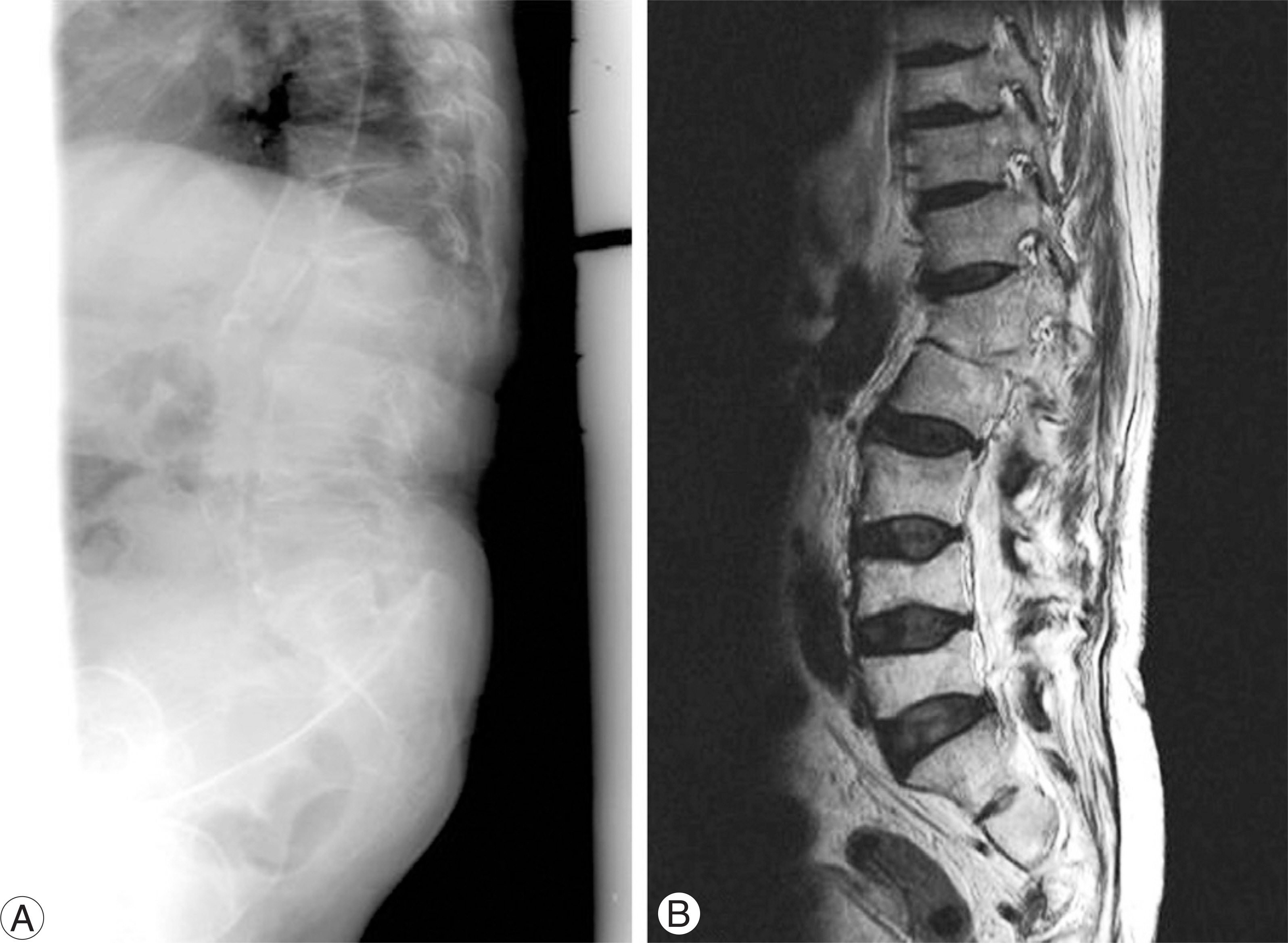
Fig. 1. Radiography (A) and MRI (B) of the thoracolumbar spine taken before injury showed acute angular kyphosis following tuberculous spondylitis.
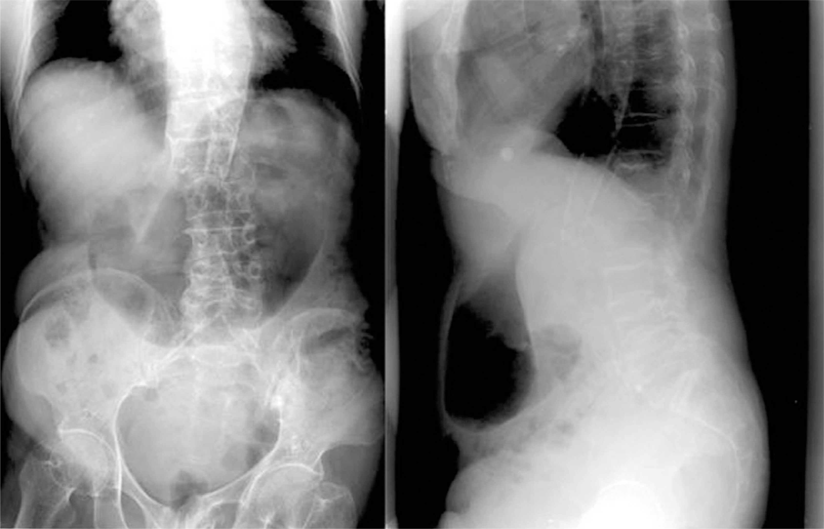
Fig. 2. Preoperative radiography after fall down showed spinal fracture through the fused vertebral body following tuberculous spondylitis between T12 and L2, which resulted in spontaneous correction of kyphosis.
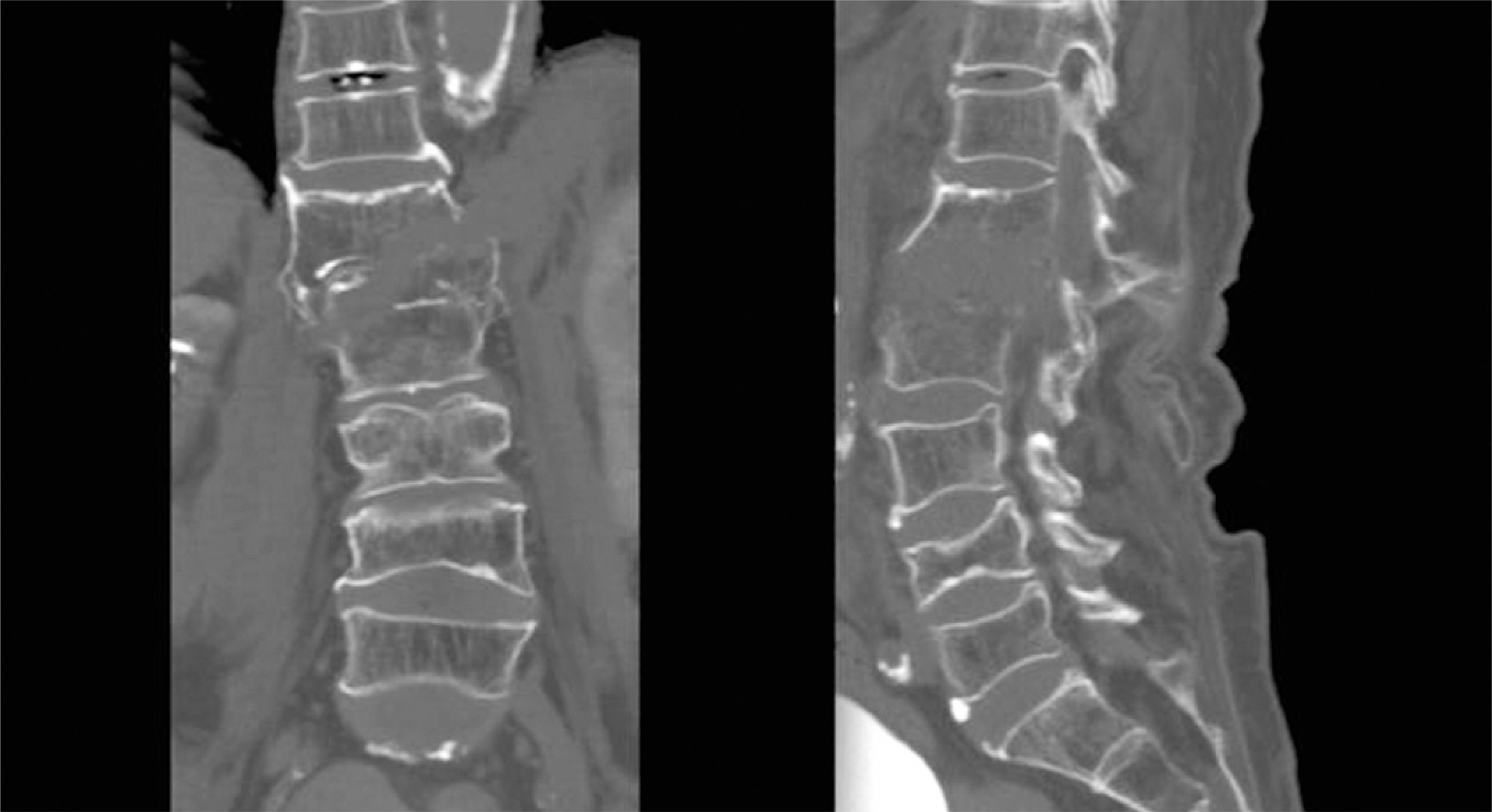
Fig. 3. Preoperative CT scan showed bony gap and oblique fracture from right side to left side and lateral displacement.
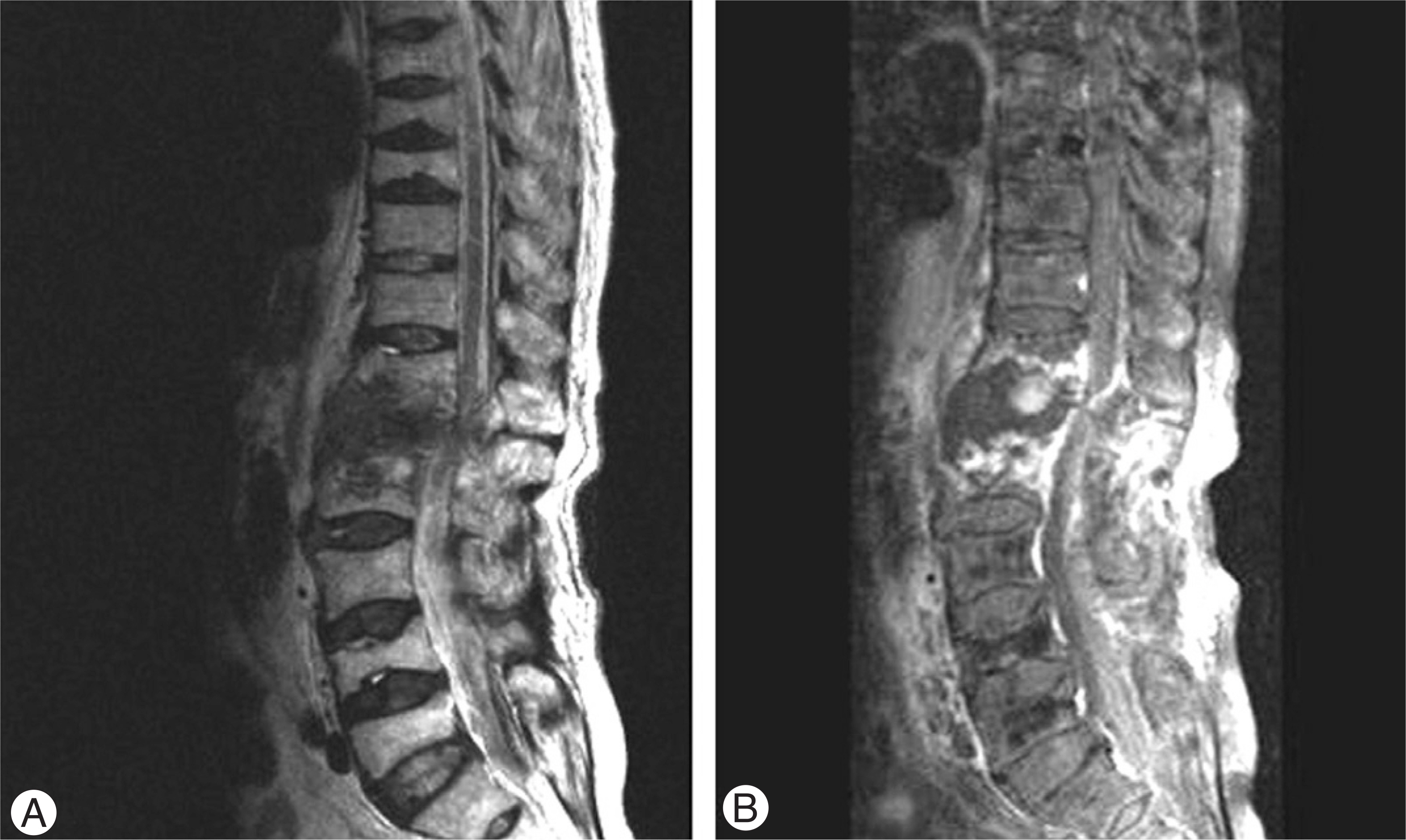
Fig. 4. T2-weighted sagittal MR image (A) showed hematoma with mixed signal intensity but no definitive cord discontinuity. Gadolinium enhanced sagittal MR image (B) showed well defined irregular shape hematoma with enhancing lesion.
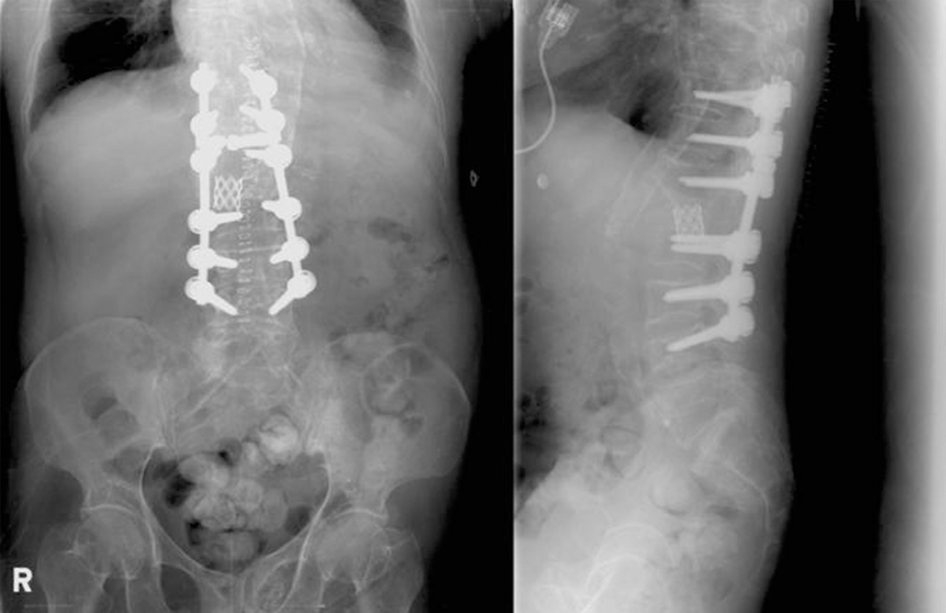
Fig. 5. Postoperative plain radiography showed significant improvement of the kyphotic deformity and sagittal balance.
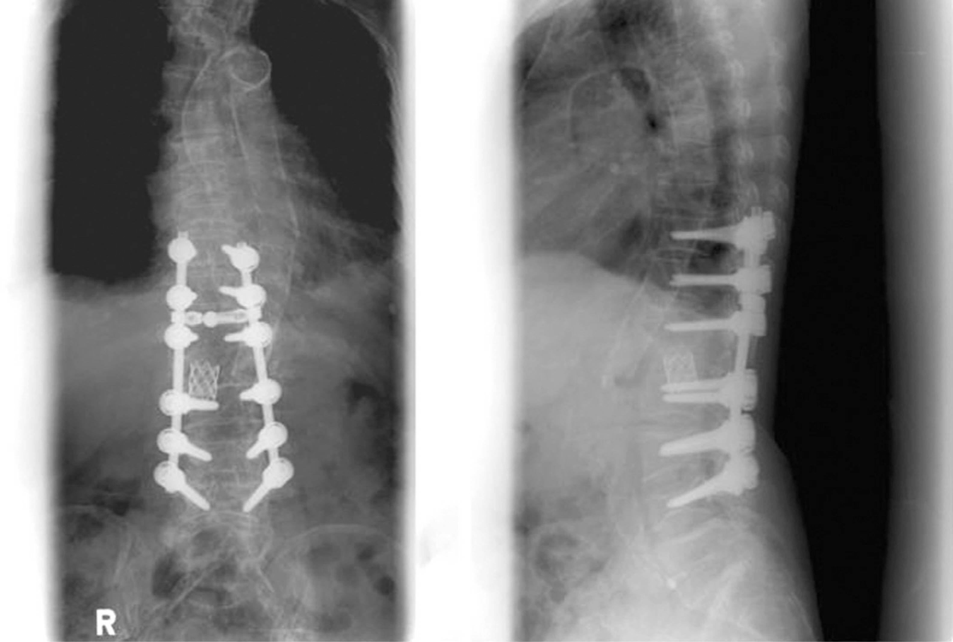
Fig. 6. The patient was doing well after one year followup and radiography showed well-maintained kyphotic angle.
Reference
-
1). Suk SI. Spinal surgery 2nd ed. Seoul. Newest Medical Publishing Co;p. 423. 2004.2). Lonstein JE. Cord compression. (in. Bradford DS, editor. Moe’ textbook of scoliosis and other spinal deformities. 3rd. Philadelphia: WB saunders Co;p. 534–540. 1995.3). Leatherman KD, Dickson RA. Two-stage corrective surgery for congenital deformities of the spine. J Bone Joint Surg Br. 1979; 61:324–328.
Article4). Bradford DS, Boachie-Adjei O. One-stage anterior and posterior hemivertebral resection and arthrodesis for congenital scoliosis. J Bone Joint Surg Am. 1990; 72:536–540.
Article5). Bradford DS, Trbus CB. Vertebral column resection for the treatment of rigid conronal decompensation. Spine. 1997; 22:1590–1599.6). Suk SI, Kim JH, Lee SM, Chung ER, Lee JH. Anterior-posterior surgery versus posterior closing wedge osteotomy in posttraumatic kyphosis with neurologic compromised osteoporotic fracture. Spine. 2003; 28:2170–2175.
Article7). Suk SI, Kim JH, Kim WJ, Lee SM, Chung ER, Nah KH. Posterior vertebral column resection for severe spinal deformities. Spine. 2002; 27:2374–2382.
Article
- Full Text Links
-
- Actions
-
Cited
- CITED
-
- Close
- Share
-
- Similar articles
-
- A new Technique of Posterior Closing Apical Correctional Osteotomy of the Thoracic of Lumbar Spine: A Report of Three Cases
- The correction of spinal deformities due to Tuberculous Spondylitis: 6 cases Report
- Correction of Kyphosis Caused by Tuberculosis of the Spine
- A Study on the Change of the Kyphosis of the Tuberculous Spine in Children following Ambulatory Treatment (II. Kyphosis and Pulmonary Function)
- Paraplegia in an Ankylosing Spondylitis Patient with a Neglected Spine Fracture after Osteosynthesis for Fracture of the Femur: A Case Report
