The Growth of an Extrapancreatic Solid Pseudopapillary Tumor from the Greater Omentum: A Case Report
- Affiliations
-
- 1Department of Radiology, Wonkwang University Hospital, Korea. yjyh@wonkwang.ac.kr
- 2Department of Pathology, Wonkwang University Hospital, Korea.
- KMID: 2208997
- DOI: http://doi.org/10.3348/jksr.2010.62.1.67
Abstract
- A solid pseudopapillary tumor is an uncommon tumor of the pancreas that rarely metastasizes to other organs and usually shows good prognosis. An extrapancreatic tumor arising from a solid pseudopapillary tumor is very rare. We report a case of an atypical extrapancreatic solid pseudopapillary tumor that arose from the great omentum and disseminated to the peritoneum, and discuss the radiologic findings, including the CT, US, and MRI.
MeSH Terms
Figure
-
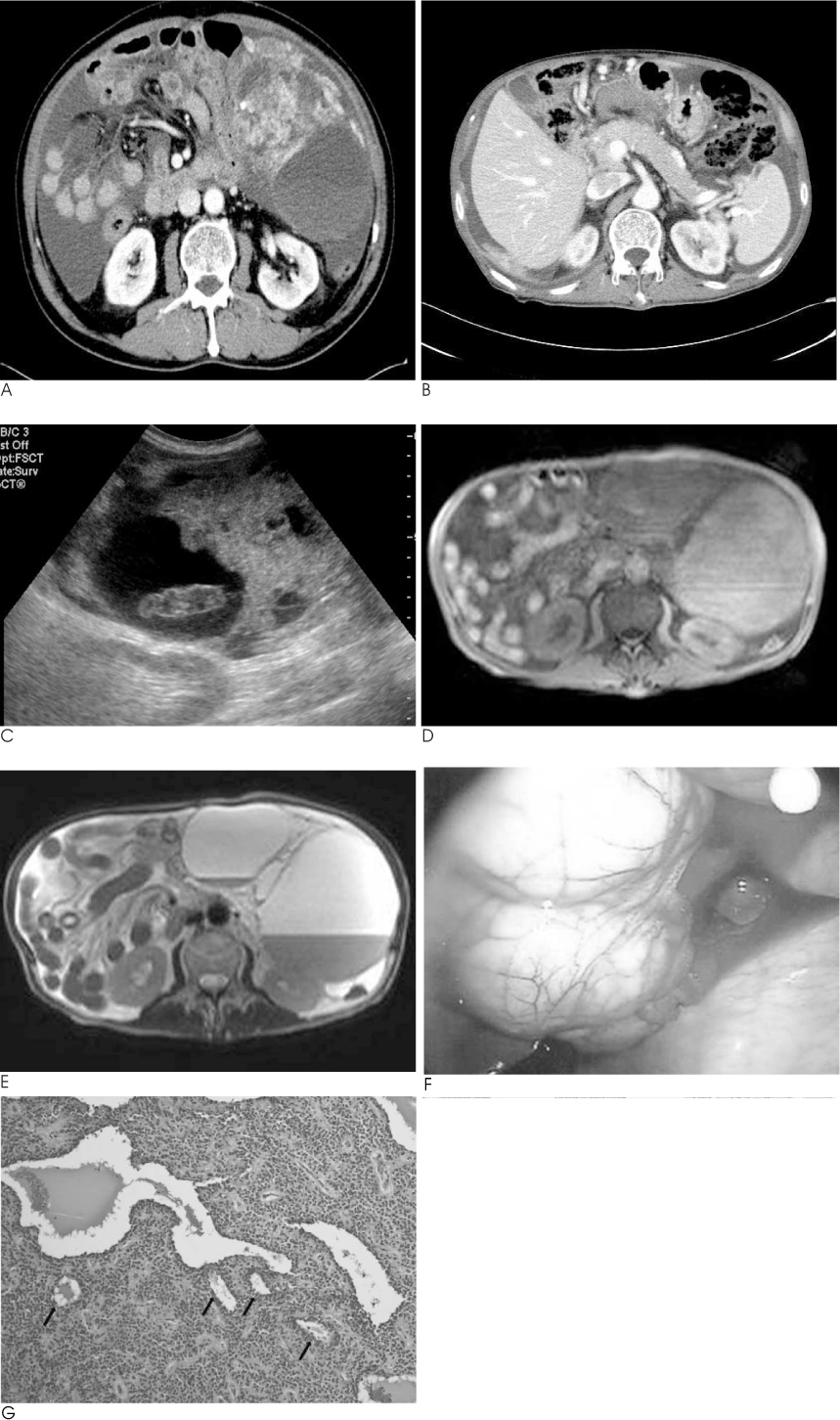
Fig. 1 A 71-year-old man with peritoneal solid pseudopapillary tumor arose at the greater omentum. A, B. Contrast enhanced axial CT images show a large complex mass at left upper quadrant, with mixed cystic and solid nature, and slight contrast enhancement of peripheral solid portion of the mass. There is no mass at pancreas. Multiple enhancing nodules at right subphrenic space with large amount of ascite. C. Abdominal US shows a complex mass with hyperechoic solid portion and anechoic cystic portions at LUQ area. D, E. MR images show that the mass has heterogeneous internal signal intensity, which indicates that the mass is more complex than suggested by the CT findings. Axial T1-weighted image (D) shows homogenous high signal intensity of the cystic mass and T2-weighted MR image (E) shows that the mass has fluid-fluid level, a finding consistent with hemorrhage. F. Photograph of peritoneal mass at greater omentum in the laparoscopic biopsy. G. Micro-photogram shows multifocal cystic changes (arrows) and scanty microvasculature in monomorphic solid pattern of tumor tissue (hematoxylin-eosin, original magnification ×100).
Reference
-
1. Coleman KM, Doherty MC, Bigler SA. Solid pseudopapillary tumor of the pancreas. Radiographics. 2003; 23:1644–1648.2. Choi JY, Kim MJ, Kim JH, Kim SH, Lim JS, Oh YT, et al. Solid pseudopapillary tumor of the pancreas: typical and atypical manifestations. AJR Am J Roentgenol. 2006; 187:W178–W186.3. Hibi T, Ojima H, Sakamoto Y, Kosuge T, Shimada K, Sano T, et al. A solid pseudopapillary tumor arising from the greater omentum followed by multiple metastases with increasing malignant potential. J Gastroenterol. 2006; 41:276–281.4. Fukunaga M. Pseudopapillary solid cystic tumor arising from an extrapancreatic site. Arch Pathol Lab Med. 2001; 125:1368–1371.5. Ishikawa O, Ishiguro S, Ohhigashi H, Sasaki Y, Yasuda T, Imaoka S, et al. Solid and papillary neoplasm arising from an ectopic pancreas in the mesocolon. Am J Gastroenterol. 1990; 85:597–601.6. Kim YI, Kim ST, Lee GK, Choi BI. Papillary cystic tumor of the liver. A case report with ultrastructural observation. Cancer. 1990; 65:2740–2746.7. Klöppel G, Maurer R, Hofmann E, Luthold K, Oscarson J, Forsby N, et al. Solid-cystic (papillary-cystic) tumours within and outside the pancreas in men: report of two patients. Virchows Arch A Pathol Anat Histopathol. 1991; 418:179–183.8. Kloppel G, Solcia E, Longnecker DS, Capella C, Sobin LH. World Health Organization: histological typing of tumours of the exocrine pancreas. 2nd. Berlin: Springer-Verlag;1996.9. Lam KY, Lo CY, Fan ST. Pancreatic solid-cystic-papillary tumor: clinicopathologic features in eight patients from Hong Kong and review of the literature. World J Surg. 1999; 23:1045–1050.10. Adsay NV, Klimstra DS. Cystic forms of typically solid pancreatic tumors. Semin Diagn Pathol. 2000; 17:81–88.
- Full Text Links
-
- Actions
-
Cited
- CITED
-
- Close
- Share
-
- Similar articles
-
- Radiologic Findings in Extrapancreatic Solid Pseudopapillary Tumor with Aggressive Behavior: a Case Report
- Solid Pseudopapillary Tumor of the Pancreas in Child: A Case Report
- A Case of Atypical Solid-pseudopapillary Tumor of the Pancreas
- A Case of Primary Omental Leiomyosarcoma
- A Case of Solid Pseudopapillary Tumor of the Pancreas Presenting as a Submucosal Tumor with Hemorrhage
