Radiologic Findings in Extrapancreatic Solid Pseudopapillary Tumor with Aggressive Behavior: a Case Report
- Affiliations
-
- 1Department of Radiology, Chung-Ang University Hospital, Chung-Ang University College of Medicine, Seoul, Korea. seolly1024@gmail.com
- 2Department of Pathology, Soonchunhyang University Cheonan Hospital, Soonchunhyang University College of Medicine, Cheonan, Korea.
- KMID: 2396390
- DOI: http://doi.org/10.3346/jkms.2017.32.12.2079
Abstract
- Solid pseudopapillary tumor (SPT) is a low grade malignant tumor in the pancreas, and extrapancreatic SPT is extremely rare. We report a case of a 61-year-old woman who complained abdominal pain with diffuse tenderness. She was diagnosed with extrapancreatic SPT with extensive peritoneal dissemination and hepatic metastases. Although a few cases have reported imaging findings of extrapancreatic SPT, there have been no reports of extrapancreatic SPT with aggressive tumor behavior and dismal prognosis. Although imaging features closely resembled those of classical pancreatic SPTs, malignant transformation of extrapancreatic SPT should be considered when focal discontinuity of the tumor capsule with ill-defined margin and invasion of adjacent structures were identified.
Figure
-
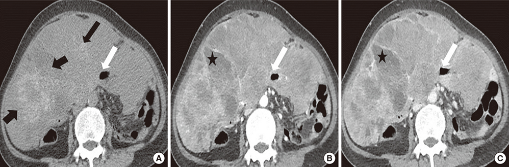
Fig. 1 CT images in mid-abdomen level. (A) Precontrast, (B) portal venous phase, (C) 3-minute delayed phase. The tumor shows heterogeneously persistent enhancement, which replaces entire abdomen. Amorphous calcifications (black arrows) and internal necrotic foci (asterisks) are also identified. Despite of bowel invasion (white arrows), bowel obstruction does not occur. CT = computed tomography.
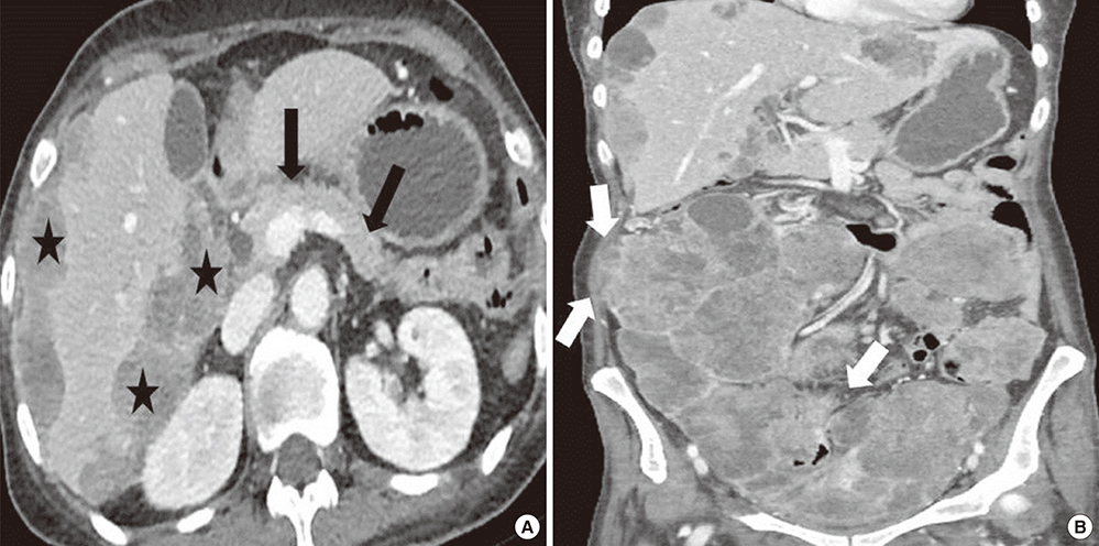
Fig. 2 CT images of SPT on portal venous phase with adjacent organ invasions. (A) Axial image, (B) coronal image. Heterogeneously enhanced seeding masses (asterisks) scallop the liver surface. The masses are separated from pancreatic head. Pancreas (black arrows) is normal. Focally ill-defined margin of the mass reveals direct invasion to right abdominal wall (white arrows). CT = computed tomography, SPT = solid pseudopapillary tumor.
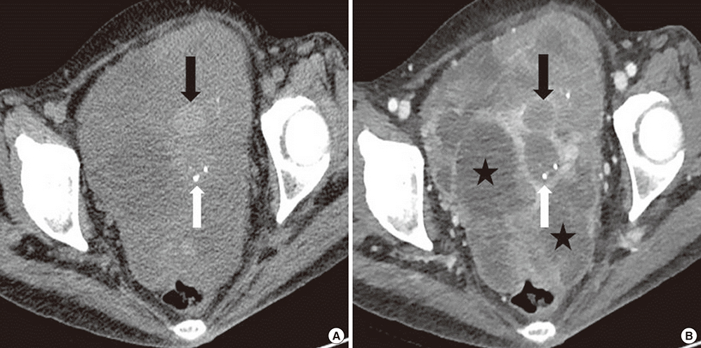
Fig. 3 CT images in the pelvic cavity level. (A) Precontrast, (B) portal venous phase. The tumor shows intralesional hemorrhage (black arrows), scattered calcification (white arrow) and internal necrotic foci (asterisks). Both ovaries are not visible. CT = computed tomography.

Fig. 4 Transverse ultrasonographic images of the SPT. (A, B) Gray scalses, (C) color Doppler image. The tumor shows well-encapsulated, heterogeneous echogenicity in gray-scale images (A, B). Cystic or necrotic components (asterisks), internal echogenic spots (white arrows), and septations (short black arrows) are identified. The posterior capsule of the mass (black arrows) shows an echogenic rim with good through-transmission of sound. Color Doppler image (C) shows increased vascularity at the solid portion of the tumor. Curved arrow indicates biopsy needle. SPT = solid pseudopapillary tumor.
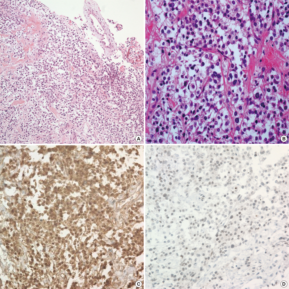
Fig. 5 Histological features of the tumor. (A) H & E stained, × 100, (B) H & E stained, × 400. (C) Immunohistochemical staining for β-catenin nucleus, (D) Immunohistochemical staining for E-cadherin. The tumor (A, B) shows solid and pseudopapillary growth with a monotonous round to oval nucleus, fine chromatin and abundant mitosis. Translocation of β-catenin nucleus (C) and loss of E-cadherin (D) are observed on immunohistochemical staining. H & E = hematoxylin and eosin.
Reference
-
1. Hibi T, Ojima H, Sakamoto Y, Kosuge T, Shimada K, Sano T, Sakamoto M, Kitajima M, Yamasaki S. A solid pseudopapillary tumor arising from the greater omentum followed by multiple metastases with increasing malignant potential. J Gastroenterol. 2006; 41:276–281.2. Papavramidis T, Papavramidis S. Solid pseudopapillary tumors of the pancreas: review of 718 patients reported in English literature. J Am Coll Surg. 2005; 200:965–972.3. Ishikawa O, Ishiguro S, Ohhigashi H, Sasaki Y, Yasuda T, Imaoka S, Iwanaga T, Nakaizumi A, Fujita M, Wada A. Solid and papillary neoplasm arising from an ectopic pancreas in the mesocolon. Am J Gastroenterol. 1990; 85:597–601.4. Tornóczky T, Kálmán E, Jáksó P, Méhes G, Pajor L, Kajtár GG, Battyány I, Davidovics S, Sohail M, Krausz T. Solid and papillary epithelial neoplasm arising in heterotopic pancreatic tissue of the mesocolon. J Clin Pathol. 2001; 54:241–245.5. Kim YI, Kim ST, Lee GK, Choi BI. Papillary cystic tumor of the liver. A case report with ultrastructural observation. Cancer. 1990; 65:2740–2746.6. Deshpande V, Oliva E, Young RH. Solid pseudopapillary neoplasm of the ovary: a report of 3 primary ovarian tumors resembling those of the pancreas. Am J Surg Pathol. 2010; 34:1514–1520.7. Walter T, Hommell-Fontaine J, Hervieu V, Adham M, Poncet G, Dumortier J, Lombard-Bohas C, Scoazec JY. Primary malignant solid pseudopapillary tumors of the gastroduodenal area. Clin Res Hepatol Gastroenterol. 2011; 35:227–233.8. Slidell MB, Schmidt EF, Jha RC, Rossi CT, Becker TE, Guzzetta PC. Solid pseudopapillary tumor in a pancreatic rest of the jejunum. J Pediatr Surg. 2009; 44:E25–E27.9. Miyazaki Y, Miyajima A, Maeda T, Yuge K, Hasegawa M, Kosaka T, Kikuchi E, Kameyama K, Jinzaki M, Nakagawa K, et al. Extrapancreatic solid pseudopapillary tumor: case report and review of the literature. Int J Clin Oncol. 2012; 17:165–168.10. Zhu H, Xia D, Wang B, Meng H. Extrapancreatic solid pseudopapillary neoplasm: report of a case of primary retroperitoneal origin and review of the literature. Oncol Lett. 2013; 5:1501–1504.11. Junzu G, Yanbin S, Suxia W, Janjun D. A case of extrapancreatic solid pseudopapillary tumor in the retroperitoneum. Jpn J Radiol. 2012; 30:598–601.12. Kim MJ, Jang SJ, Yu E. Loss of E-cadherin and cytoplasmic-nuclear expression of β-catenin are the most useful immunoprofiles in the diagnosis of solid-pseudopapillary neoplasm of the pancreas. Hum Pathol. 2008; 39:251–258.13. Choi JY, Kim MJ, Kim JH, Kim SH, Lim JS, Oh YT, Chung JJ, Yoo HS, Lee JT, Kim KW. Solid pseudopapillary tumor of the pancreas: typical and atypical manifestations. AJR Am J Roentgenol. 2006; 187:W178–W186.14. Yin Q, Wang M, Wang C, Wu Z, Yuan F, Chen K, Tang Y, Zhao X, Miao F. Differentiation between benign and malignant solid pseudopapillary tumor of the pancreas by MDCT. Eur J Radiol. 2012; 81:3010–3018.15. Lee DH, Yi BH, Lim JW, Ko YT. Sonographic findings of solid and papillary epithelial neoplasm of the pancreas. J Ultrasound Med. 2001; 20:1229–1232.16. Jung WS, Kim JK, Yu JS, Kim JH, Cho ES, Chung JJ. Comparison of abdominal ultrasonographic findings with endoscopic ultrasonographic findings of solid pseudopapillary neoplasms of the pancreas. Ultrasound Q. 2014; 30:173–178.17. Levy AD, Arnáiz J, Shaw JC, Sobin LH. From the archives of the AFIP: primary peritoneal tumors: imaging features with pathologic correlation. Radiographics. 2008; 28:583–607.
- Full Text Links
-
- Actions
-
Cited
- CITED
-
- Close
- Share
-
- Similar articles
-
- The Growth of an Extrapancreatic Solid Pseudopapillary Tumor from the Greater Omentum: A Case Report
- Solid Pseudopapillary Tumor of the Pancreas in Child: A Case Report
- A Case of Atypical Solid-pseudopapillary Tumor of the Pancreas
- An Unusual Presentation of a Solid Pseudopapillary Tumor of the Pancreas Mimicking Adenocarcinoma
- A Case of Solid Pseudopapillary Tumor of the Pancreas Presenting as a Submucosal Tumor with Hemorrhage
